| [1] |
Amier RP, Tijssen RYG, Teunissen PFA, et al. Predictors of intramyocardial hemorrhage after reperfused ST-segment elevation myocardial infarction. J Am Heart Assoc 2017; 6(8):e005651. doi: 10.1161/jaha.117.005651.
doi: 10.1161/jaha.117.005651
|
| [2] |
Nijveldt R, Beek AM, Hirsch A, et al. Functional recovery after acute myocardial infarction: comparison between angiography, electrocardiography, and cardiovascular magnetic resonance measures of microvascular injury. J Am Coll Cardiol 2008; 52(3):181-9. doi: 10.1016/j.jacc.2008.04.006.
doi: 10.1016/j.jacc.2008.04.006
pmid: 18617066
|
| [3] |
Schwaiger JP, Reinstadler SJ, Tiller C, et al. Baseline LV ejection fraction by cardiac magnetic resonance and 2D echocardiography after ST-elevation myocardial infarction—influence of infarct location and prognostic impact. Eur Radiol 2020; 30(1):663-71. doi: 10.1007/s00330-019-06316-3.
doi: 10.1007/s00330-019-06316-3
pmid: 31428825
|
| [4] |
Amzulescu MS, De Craene M, Langet H, et al. Myocardial strain imaging: review of general principles, validation, and sources of discrepancies. Eur Heart J Cardiovasc Imaging 2019; 20(6):605-19. doi: 10.1093/ehjci/jez041.
doi: 10.1093/ehjci/jez041
pmid: 30903139
|
| [5] |
Wamil M, Borlotti A, Liu D, et al. Combined T1-mapping and tissue tracking analysis predicts severity of ischemic injury following acute STEMI—an Oxford Acute Myocardial Infarction (OxAMI) study. Int J Cardiovasc Imaging 2019; 35(7):1297-308. doi: 10.1007/s10554-019-01542-8.
doi: 10.1007/s10554-019-01542-8
|
| [6] |
Khan JN, Singh A, Nazir SA, et al. Comparison of cardiovascular magnetic resonance feature tracking and tagging for the assessment of left ventricular systolic strain in acute myocardial infarction. Eur J Radiol 2015; 84(5):840-8. doi: 10.1016/j.ejrad.2015.02.002.
doi: 10.1016/j.ejrad.2015.02.002
pmid: 25743248
|
| [7] |
Everaars H, Robbers L, Götte M, et al. Strain analysis is superior to wall thickening in discriminating between infarcted myocardium with and without microvascular obstruction. Eur Radiol 2018; 28(12):5171-81. doi: 10.1007/s00330-018-5493-0.
doi: 10.1007/s00330-018-5493-0
pmid: 29948065
|
| [8] |
Fischer K, Linder OL, Erne SA, et al. Reproducibility and its confounders of CMR feature tracking myocardial strain analysis in patients with suspected myocarditis. Eur Radiol 2022; 32(5):3436-46. doi: 10.1007/s00330-021-08416-5.
doi: 10.1007/s00330-021-08416-5
|
| [9] |
Eitel I, Stiermaier T, Lange T, et al. Cardiac magnetic resonance myocardial feature tracking for optimized prediction of cardiovascular events following myocardial infarction. JACC Cardiovasc Imaging 2018; 11(10):1433-44. doi: 10.1016/j.jcmg.2017.11.034.
doi: S1936-878X(17)31176-2
pmid: 29454776
|
| [10] |
Gavara J, Rodriguez-Palomares JF, Valente F, et al. Prognostic value of strain by tissue tracking cardiac magnetic resonance after ST-segment elevation myocardial infarction. JACC Cardiovasc Imaging 2018; 11(10):1448-57. doi: 10.1016/j.jcmg.2017.09.017.
doi: S1936-878X(17)30985-3
pmid: 29248649
|
| [11] |
O'Regan DP, Ariff B, Baksi AJ, et al. Salvage assessment with cardiac MRI following acute myocardial infarction underestimates potential for recovery of systolic strain. Eur Radiol 2013; 23(5):1210-7. doi: 10.1007/s00330-012-2715-8.
doi: 10.1007/s00330-012-2715-8
pmid: 23179525
|
| [12] |
Reindl M, Tiller C, Holzknecht M, et al. Global longitudinal strain by feature tracking for optimized prediction of adverse remodeling after ST-elevation myocardial infarction. Clin Res Cardiol 2021; 110(1):61-71. doi: 10.1007/s00392-020-01649-2.
doi: 10.1007/s00392-020-01649-2
|
| [13] |
Kidambi A, Mather AN, Swoboda P, et al. Relationship between myocardial edema and regional myocardial function after reperfused acute myocardial infarction: an MR imaging study. Radiology 2013; 267(3):701-8. doi: 10.1148/radiol.12121516.
doi: 10.1148/radiol.12121516
pmid: 23382292
|
| [14] |
Zhao H, Lee AP, Li Z, et al. Impact of intramyocardial hemorrhage and microvascular obstruction on cardiac mechanics in reperfusion injury: a speckle-tracking echocardiographic study. J Am Soc Echocardiogr 2016; 29(10):973-82. doi: 10.1016/j.echo.2016.06.011.
doi: 10.1016/j.echo.2016.06.011
|
| [15] |
Thygesen K, Alpert JS, Jaffe AS, et al. Third universal definition of myocardial infarction. J Am Coll Cardiol 2012; 60(16):1581-98. doi: 10.1016/j.jacc.2012.08.001.
doi: 10.1016/j.jacc.2012.08.001
pmid: 22958960
|
| [16] |
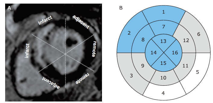
Cerqueira MD, Weissman NJ, Dilsizian V, et al. Standardized myocardial segmentation and nomenclature for tomographic imaging of the heart. A statement for healthcare professionals from the Cardiac Imaging Committee of the Council on Clinical Cardiology of the American Heart Association. Circulation 2002; 105 (4):539-42. doi: 10.1161/hc0402.102975.
doi: 10.1161/hc0402.102975
pmid: 11815441
|
| [17] |
Zou Q, Zheng T, Zhou SL, et al. Quantitative evaluation of myocardial strain after myocardial infarction with cardiovascular magnetic resonance tissue-tracking imaging. Int Heart J 2020; 61(3):429-36. doi: 10.1536/ihj.19-384.
doi: 10.1536/ihj.19-384
pmid: 32350202
|
| [18] |
Bulluck H, Carberry J, Carrick D, et al. A noncontrast CMR risk score for long-term risk stratification in reperfused ST-segment elevation myocardial infarction. JACC Cardiovasc Imaging 2022; 15(3):431-40. doi: 10.1016/j.jcmg.2021.08.006.
doi: 10.1016/j.jcmg.2021.08.006
pmid: 35272808
|
| [19] |
He J, Yang W, Wu W, et al. Early diastolic longitudinal strain rate at MRI and outcomes in heart failure with preserved ejection fraction. Radiology 2022; 302(1):E5. doi: 10.1148/radiol.2021219026.
doi: 10.1148/radiol.2021219026
pmid: 34928734
|
| [20] |
Lee JW, Hur JH, Yang DH, et al. Guidelines for cardiovascular magnetic resonance imaging from the Korean Society of Cardiovascular Imaging-Part 2: interpretation of cine, flow, and angiography data. Korean J Radiol 2019; 20(11):1477-90. doi: 10.3348/kjr.2019.0407.
doi: 10.3348/kjr.2019.0407
pmid: 31606953
|
| [21] |
Carrick D, Haig C, Ahmed N, et al. Myocardial hemorrhage after acute reperfused ST-segment-elevation myocardial infarction: relation to microvascular obstruction and prognostic significance. Circ Cardiovasc Imaging 2016; 9(1):e004148. doi: 10.1161/circimaging.115.004148.
doi: 10.1161/circimaging.115.004148
|
| [22] |
Huang Y, Lei D, Chen Z, et al. Factors associated with microvascular occlusion in patients with ST elevation myocardial infarction after primary percutaneous coronary intervention. J Int Med Res 2021; 49(6): 3000605211024490. doi: 10.1177/03000605211024490.
doi: 10.1177/03000605211024490
|
| [23] |
Podlesnikar T, Pizarro G, Fernández-Jiménez R, et al. Left ventricular functional recovery of infarcted and remote myocardium after ST-segment elevation myocardial infarction (METOCARD-CNIC randomized clinical trial substudy). J Cardiovasc Magn Reson 2020; 22: (1):44. doi: 10.1186/s12968-020-00638-8.
doi: 10.1186/s12968-020-00638-8
|
| [24] |
Lange T, Stiermaier T, Backhaus SJ, et al. Functional and prognostic implications of cardiac magnetic resonance feature tracking-derived remote myocardial strain analyses in patients following acute myocardial infarction. Clin Res Cardiol 2021; 110(2):270-80. doi: 10.1007/s00392-020-01747-1.
doi: 10.1007/s00392-020-01747-1
pmid: 33083869
|
| [25] |
Zhang L, Mandry D, Chen B, et al. Impact of microvascular obstruction on left ventricular local remodeling after reperfused myocardial infarction. J Magn Reson Imaging 2018; 47(2):499-510. doi: 10.1002/jmri.25780.
doi: 10.1002/jmri.25780
pmid: 28580619
|
| [26] |
Pankaj G, Ananth K, James RJ, et al. Ventricular longitudinal function is associated with microvascular obstruction and intramyocardial haemorrhage. Open Heart 2016; 3(1): e000337. doi: 10.1136/openhrt-2015-000337.
doi: 10.1136/openhrt-2015-000337
|
| [27] |
Claus P, Omar AMS, Pedrizzetti G, et al. Tissue tracking technology for assessing cardiac mechanics: principles, normal values, and clinical applications. JACC Cardiovasc Imaging 2015; 8(12):1444-60. doi: 10.1016/j.jcmg.2015.11.001.
doi: S1936-878X(15)00845-1
pmid: 26699113
|
| [28] |
Regenfus M, Schlundt C, Krähner R, et al. Six-year prognostic value of microvascular obstruction after reperfused ST-elevation myocardial infarction as assessed by contrast-enhanced cardiovascular magnetic resonance. Am J Cardiol 2015; 116(7):1022-7. doi: 10.1016/j.amjcard2015.06.034.
doi: 10.1016/j.amjcard.2015.06.034
pmid: 26260397
|
| [29] |
Smiseth OA, Torp H, Opdahl A, et al. Myocardial strain imaging: how useful is it in clinical decision making? Eur Heart J 2016; 37(15):1196-207. doi: 10.1093/eurheartj/ehv529.
doi: 10.1093/eurheartj/ehv529
pmid: 26508168
|
 )
)