Chinese Medical Sciences Journal ›› 2018, Vol. 33 ›› Issue (3): 188-193.doi: 10.24920/31804

• 病例报告 • 上一篇    下一篇

多参数磁共振成像诊断小脑血管母细胞瘤1例报告

陈志晔1,2,3,刘梦琦1,2,于生元3,马林2,*()   

  1. 1 解放军总医院海南分院放射科,三亚 572013
    2 解放军总医院放射科,北京 100853
    3 神经内科,北京 100853
  • 出版日期:2018-09-30 发布日期:2018-07-16
  • 通讯作者: 马林 E-mail:cjr.malin@vip.163.com

Multi-parametric MRI Diagnoses Cerebellar Hemangioblastoma: A Case Report

Chen Zhiye1,2,3,Liu Mengqi1,2,Yu Shengyuan3,Ma Lin2,*()   

  1. 1 Department of Radiology, Hainan Branch of Chinese PLA General Hospital, Sanya, Hainan 572013, China
    2 Department of Radiology, Chinese PLA General Hospital, Beijing 100853, China
    3 Department of Neurology, Chinese PLA General Hospital, Beijing 100853, China
  • Published:2018-09-30 Online:2018-07-16
  • Contact: Ma Lin E-mail:cjr.malin@vip.163.com

摘要:

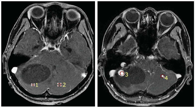
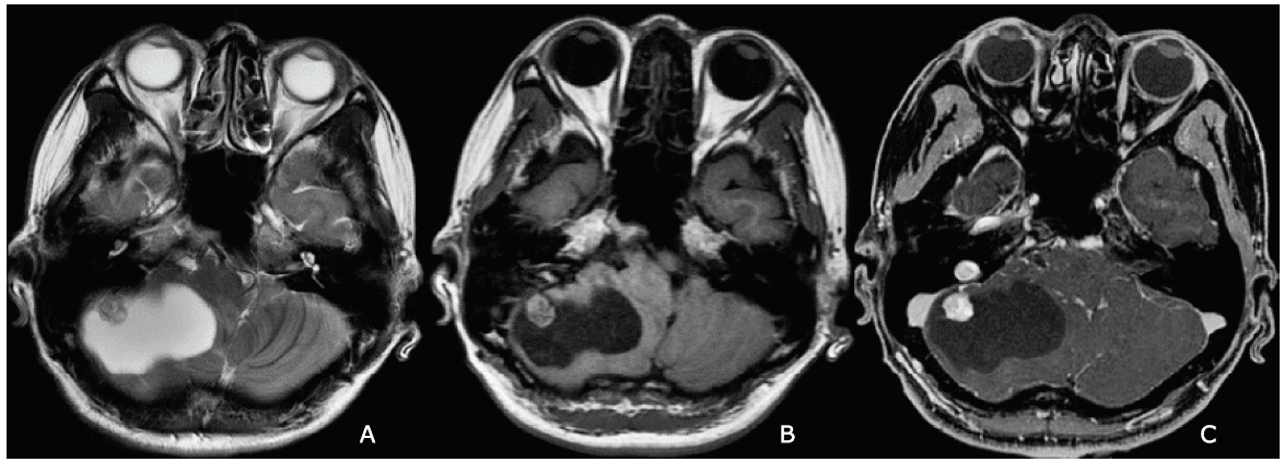
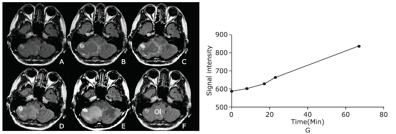
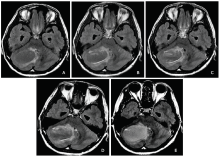
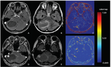
本研究采用对比剂增强T2液体衰减翻转恢复成像及动态对比剂增强磁共振成像描述1例小脑血管母细胞瘤的影像学表现。T2液体衰减翻转恢复成像显示右侧小脑可见一囊性病变并壁结节形成。注射对比剂5.6~23分钟后,病变囊壁在T2液体衰减翻转恢复成像上明显强化,动态对比剂增强磁共振成像证实囊壁及壁结节均较对侧正常小脑表现为高Ktrans、Kep及Ve值。注射对比剂后,囊性成分在T2液体衰减翻转恢复成像表现为渐进性强化,在67分钟时达到最高信号强度。总之,T2液体衰减翻转恢复成像上囊壁早期强化可能是囊性血管母细胞瘤的特征性影像学表现,有助于血管母细胞瘤的术前早期诊断。

关键词: 动态对比剂增强磁共振成像, 液体翻转恢复成像, 血管母细胞瘤, 诊断

Abstract:

We performed contrast-enhanced T2 fluid-attenuated inversion recovery (T2-FLAIR) and dynamic contrast enhanced MRI to illustrate the imaging characteristics of one case of hemangioblastoma. T2-FLAIR showed a large cyst located in the right cerebellum with mural nodule. The intensely enhancing cyst wall was observed on enhanced T2-FLAIR images acquired from 5.6 to 23 minutes after contrast administration, and quantitative dynamic contrast enhanced-MRI demonstrated that both the cyst wall and mural nodule presented high Ktrans, Kep and Ve values compared with the contralateral normal cerebellar tissues. The cyst showed gradual enhancement and reached the highest signal intensity at 67 minutes after contrast administration on enhanced T2-FLAIR images. In conclusion, early enhancement of cyst wall on T2-FLAIR might be the characteristic imaging findings for cystic hemangioblastoma, which may assist in the diagnosis of hemangioblastoma preoperatively.

Key words: dynamic contrast enhanced MRI, fluid-attenuated inversion recovery, hemangioblastoma, diagnosis

Copyright © 2018 Chinese Academy of Medical Sciences. All right reserved.
 
www.cmsj.cams.cn
京公安备110402430088 京ICP备06002729号-1  Powered by Magtech.

Supervised by National Health Commission of the People's Republic of China

9 Dongdan Santiao, Dongcheng district, Beijing, 100730 China

Tel: 86-10-65105897  Fax:86-10-65133074 

E-mail: cmsj@cams.cn  www.cmsj.cams.cn

Copyright © 2018 Chinese Academy of Medical Sciences

All right reserved.

京公安备110402430088  京ICP备06002729号-1