Chinese Medical Sciences Journal ›› 2019, Vol. 34 ›› Issue (1): 10-17.doi: 10.24920/003548
• Original Articles • Previous Articles Next Articles
Differential Diagnostic Value of Texture Feature Analysis of Magnetic Resonance T2 Weighted Imaging between Glioblastoma and Primary Central Neural System Lymphoma
Wang Botao1, Liu Mingxia2, *( ), Chen Zhiye1, 3, *()
- 1 Department of Radiology, Hainan Hospital of Chinese PLA General Hospital, Sanya, Hainan 572013, China
2 Department of Radiology, PLA 305 Hospital, Beijing 100017, China
3 Department of Radiology, Chinese PLA General Hospital, Beijing 100853, China
-
Received:2018-12-28Revised:2019-02-22Published:2019-03-30Online:2019-04-08 -
Contact:Liu Mingxia,Chen Zhiye E-mail:lvmgxx@163.com;yyqf@hotmail.com
Cite this article
Wang Botao, Liu Mingxia, Chen Zhiye. Differential Diagnostic Value of Texture Feature Analysis of Magnetic Resonance T2 Weighted Imaging between Glioblastoma and Primary Central Neural System Lymphoma[J].Chinese Medical Sciences Journal, 2019, 34(1): 10-17.
share this article
Add to citation manager EndNote|Reference Manager|ProCite|BibTeX|RefWorks
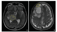
Figure 1.
A. A 33 year-old male patient with lyphoma; B. A 50 years old man with glioblastoma. The region of interest was placed on the solid part of the lesion to measure the texture parameters on the T2 weighted image."
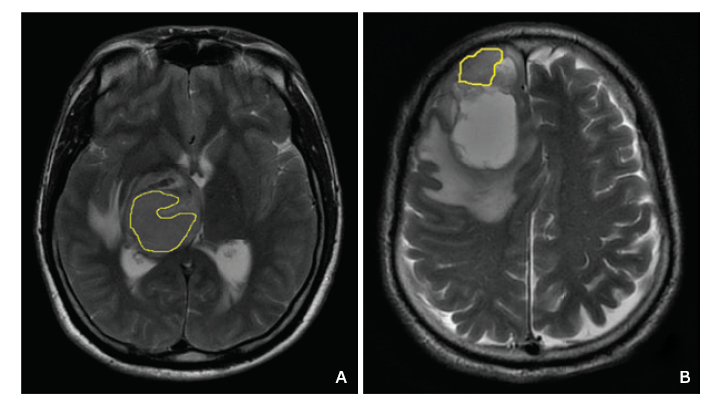
Figure 1.
Figure 2.
A. The lymphoma of the left temporal lobe presented ‘flame-like edema’; B. The glioblastoma of the right temporal lobe presented ‘Rosette’ enhancement; C. The lymphoma of the left temporal lobe and hippocampus presented ‘incision sign’ (arrow head), which pointed to the dura is aligned with the blood supply arteries and white matter fibers. "
Figure 2.
Table 1
Comparisons of the five texture parameters of DWI between 81 patients with cerebral glioblastoma and 28 patients with primary central neural system lymphoma"
| Groups | n | ASM [median (QR)]a | Contrast [median (QR)]a | Correlation [median (QR)]a | IDM (means ±SD) | Entropy (means ±SD) |
|---|---|---|---|---|---|---|
| Glioblastoma | 81 | 0.011 (0.010) | 8.302 (9.320) | 0.030 (0.020) | 0.382±0.074 | 4.933±0.434 |
| Lymphoma | 28 | 0.015 (0.010) | 4.989 (4.580) | 0.051 (0.050) | 0.449±0.085 | 4.664±0.511 |
| t value | 820.000 | 618.500 | 760.000 | 4.089 | 2.758 | |
| P value | 0.006 | 0.000 | 0.002 | 0.000 | 0.015 | |
| 95%CI | 0.011-0.013 | 7.146-10.205 | 0.032-0.044 | 0.388-0.423 | 4.733-4.913 |
Table 1
Table 2
Receiver operating characteristic curve evaluation of the texture ASM, Contrast, Correlation, Entropy, and IDM of the model of Logistic regression between cerebral glioblastoma and primary central neural system lymphoma"
| Parameters | Area under curve | Critical value | 95%CI | Sensitivity | Specificity | Above critial value | Below critical value |
|---|---|---|---|---|---|---|---|
| ASM | 0.671 | 0.015 | 0.549-0.875 | 0.483 | 0.814 | Lymphoma | Glioblastoma |
| Contrast | 0.752 | 5.267 | 0.649-0.855 | 0.802 | 0.552 | Glioblastoma | Lymphoma |
| Correlation | 0.695 | 0.050 | 0.576-0.815 | 0.517 | 0.860 | Lymphoma | Glioblastoma |
| IDM | 0.720 | 0.409 | 0.607-0.881 | 0.655 | 0.651 | Lymphoma | Glioblastoma |
| Entropy | 0.646 | 4.683 | 0.521-0.770 | 0.686 | 0.552 | Glioblastoma | Lymphoma |
Table 2

Figure 3.
ROC curve of each texture parameter and the combined texture parameters of the model of Logistic regression for glioblastoma and primary central neural system lymphoma."
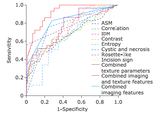
Figure 3.
| 1. |
Roth P, Stupp R, Eisele G , et al. Treatment of primary CNS lymphoma. Curr Treat Options Neurol 2014; 16(1):277. doi: 10.1007/s11940-013-0277-y.
doi: 10.1007/s11940-013-0277-y pmid: 24343307 |
| 2. |
Hervey-Jumper SL, Berger MS . Maximizing safe resection of low- and high-grade glioma. J Neurooncol 2016; 130(2):269-82. doi: 10.1007/s11060-016-2110-4.
doi: 10.1007/s11060-016-2110-4 pmid: 27174197 |
| 3. | Zhang H, Zeng WB . Study of the value of MRI in the diagnosis of high-grade gliomas. Chin J CT MRI 2017; 15(8):37-9. doi: 10.3969/j.issn.1672-5131.2017.08.011. |
| 4. | Zhang TJ, Lv S , Yue Q, et al. MRI diagnosis and differential diagnosis of intracerebral primary non-Hodgkin lymphoma. Radiol Practice 2010; 25(9):994-8. doi: 10.3969/j.issn.1000-0313.2010.09.014. |
| 5. | Cao B, Chen ZQ, Zhang JX . Comparative analysis among multimodal MRI and pathology of primary central nervous system lymphoma. Chin J CT MRI 2012; 10(6):8-11. doi: 10.3969/j.issn.1672-5131.2012.06.003. |
| 6. | Wang R, Zheng J, Du YH , et al. Value of conventional MRI features in solitary cerebral glioma grading. J Xi’an Jiaotong University (Medical Sciences) 2017; 38(6):866-71. doi: 10.7652/jdyxb201706016. |
| 7. |
Vignati A, Mazzetti S, Giannini V , et al. Texture features on T2-weighted magnetic resonance imaging: new potential biomarkers for prostate cancer aggressiveness. Phys Med Biol 2015; 60(7):2685-701. doi: 10.1088/0031-9155/60/7/2685.
doi: 10.1088/0031-9155/60/7/2685 pmid: 25768265 |
| 8. |
Chen Z, Feng F, Yang Y , et al. MR imaging findings of the corpus callosum region in the differentiation between multiple sclerosis and neuromyelitis optica. Eur J Radiol 2012; 81(11):3491-5. doi: 10.1016/j.ejrad.2012.02.010.
doi: 10.1016/j.ejrad.2012.02.010 pmid: 22445592 |
| 9. | Yu TG, Dai JZ, Feng XY . MRI and 1H-MRS characteristics of primary central nervous system lymphomas (PCNSL) . J Clin Radiol 2005; 24(8):13-7. |
| 10. |
Castellano G, Bonilha L, Li LM , et al. Texture analysis of medical images. Clin Radiol 2004; 59(12):1061-9. doi: 10.1016/j.crad.2004.07.008.
doi: 10.1016/j.crad.2004.07.008 pmid: 15556588 |
| 11. | Mohanaiah P, Sathyanarayana P, Gurukumar L . Image texture feature extraction using GLCM approach. Inter J Sci Res Publications 2014; 3(5):1-5. |
| 12. |
Wang B, Liu G, Fan W , et al. Value of texture feature analysis in the differential diagnosis of hepatic cyst and hemangioma in magnetic resonance imaging. Zhongguo Yi Xue Ke Xue Yuan Xue Bao 2017; 39(2):169-76. doi: 10.3881/j.issn.1000-503X.2017.02.002.
doi: 10.3881/j.issn.1000-503X.2017.02.002 pmid: 28483013 |
| 13. | Wang BT, He L, Liu G , et al. Value of magnetic resonance imaging texture feature analysis in the differential diagnosis between pancreatic serous cystadenoma and mucinous cystadenoma. Zhongguo Yi Xue Ke Xue Yuan Xue Bao 2018; 40(2):187-93. doi: 10.3881/j.issn.1000-503X.2018.02.008. |
| 14. | Wang BT, Liu G, He L , et al. Texture feature analysis in follow-up of pulmonary ground glass nodule. Chin J Med Imaging 2017; 25(6):441-6. doi: 10.3969/j.issn.1005-5185.2017.06.011. |
| 15. | Bo H, Ma FL, Jiao LC . Research on computation of GLCM of image texture. Dianzi Xue Bao 2006; 34(1):57-60. doi: 10.3321/j.issn:0372-2112.2006.01.032. |
| 16. |
Skogen K, Schulz A, Dormagen JB , et al. Diagnostic performance of texture analysis on MRI in grading cerebral gliomas. Eur J Radiol 2016; 85(4):824-9. doi: 10.1016/j.ejrad.2016.01.013.
doi: 10.1016/j.ejrad.2016.01.013 pmid: 26971430 |
| 17. |
Chu HH, Choi SH, Ryoo I , et al. Differentiation of true progression from pseudoprogression in glioblastoma treated with radiation therapy and concomitant temozolomide: comparison study of standard and high-b-value diffusion-weighted imaging. Radiology 2013; 269(3):831-40. doi: 10.1148/radiol.13122024.
doi: 10.1148/radiol.13122024 pmid: 23771912 |
| 18. |
Xiao DD, Yan PF, Wang YX , et al. Glioblastoma and primary central nervous system lymphoma: preoperative differentiation by using MRI-based 3D texture analysis. Clin Neurol Neurosurg 2018; 173:84-90. doi: 10.1016/j.clineuro.2018.08.004.
doi: 10.1016/j.clineuro.2018.08.004 |
| 19. |
Kunimatsu A, Kunimatsu N, Yasaka K , et al. Machine learning-based texture analysis of contrast-enhanced MR imaging to differentiate between glioblastoma and primary central nervous system lymphoma. Magn Reson Med Sci 2019; 18(1):44-52. doi: 10.2463/mrms.mp.2017-0178.
doi: 10.2463/mrms.mp.2017-0178 |
| 20. |
Suh HB, Choi YS, Bae S , et al. Primary central nervous system lymphoma and atypical glioblastoma: differentiation using radiomics approach. Eur Radiol 2018; 28(9):3832-9. doi: 10.1007/s00330-018-5368-4.
doi: 10.1007/s00330-018-5368-4 pmid: 29626238 |
| 21. |
Kunimatsu A, Kunimatsu N, Kamiya K , et al. Comparison between glioblastoma and primary central nervous system lymphoma using MR image-based texture analysis. Magn Reson Med Sci 2018; 17(1):50-7. doi: 10.2463/mrms.mp.2017-0044.
doi: 10.2463/mrms.mp.2017-0044 pmid: 28638001 |
| 22. |
Verma RK, Wiest R, Locher C , et al. Differentiating enhancing multiple sclerosis lesions, glioblastoma, and lymphoma with dynamic texture parameters analysis (DTPA): a feasibility study. Med Phys 2017; 44(8):4000-8. doi: 10.1002/mp.12356.
doi: 10.1002/mp.12356 pmid: 28543071 |
| 23. | Sun ZG, Wang XL, Zhu H , et al. The value of texture analysis in differentiating diagnosis between primary cerebral lymphoma and high-grade glioma. J Clin Radiol 2017; 36(9):1229-34. doi: 10.13437/j.cnki.jcr.2017.09.007. |
| [1] | Jian Cao, Guorong Wang, Zhiwei Wang, Zhengyu Jin. CT Texture Analysis: A Potential Biomarker for Evaluating KRAS Mutational Status in Colorectal Cancer [J]. Chinese Medical Sciences Journal, 2020, 35(4): 306-314. |
| [2] | Wang Yingwei, Zhang Xinghua, Wang Botao, Wang Ye, Liu Mengqi, Wang Haiyi, Ye Huiyi, Chen Zhiye. Value of Texture Analysis of Intravoxel Incoherent Motion Parameters in Differential Diagnosis of Pancreatic Neuroendocrine Tumor and Pancreatic Adenocarcinoma [J]. Chinese Medical Sciences Journal, 2019, 34(1): 1-9. |
| [3] | Liu Hongjuan, Zhou Huanfen, Zong Linxiong, Liu Mengqi, Wei Shihui, Chen Zhiye. MRI Histogram Texture Feature Analysis of the Optic Nerve in the Patients with Optic Neuritis [J]. Chinese Medical Sciences Journal, 2019, 34(1): 18-23. |
| [4] | Xu Jia, Wang Xuan, Jin Zhengyu, You Yan, Wang Qin, Wang Shitian, Xue Huadan. Value of Texture Analysis on Gadoxetic Acid-enhanced MR for Detecting Liver Fibrosis in a Rat Model [J]. Chinese Medical Sciences Journal, 2019, 34(1): 24-32. |
| [5] | Wang Botao, Fan Wenping, Xu Huan, Li Lihui, Zhang Xiaohuan, Wang Kun, Liu Mengqi, You Junhao, Chen Zhiye. Value of Magnetic Resonance Imaging Texture Analysis in the Differential Diagnosis of Benign and Malignant Breast Tumors [J]. Chinese Medical Sciences Journal, 2019, 34(1): 33-37. |
| [6] | Wang Guorong, Wang Zhiwei, Jin Zhengyu. Application and Progress of Texture Analysis in the Therapeutic Effect Prediction and Prognosis of Neoadjuvant Chemoradiotherapy for Colorectal Cancer [J]. Chinese Medical Sciences Journal, 2019, 34(1): 45-50. |
| [7] | Lin Lu, Feng Gu, Wei-xin Dai, Wu-yi Li, Jie Chen, Yu Xiao and Zheng-pei Zeng. Clinical and Pathological Features of Riedel‘s Thyroiditis [J]. Chinese Medical Sciences Journal, 2010, 25(3): 129-134. |
| Viewed | ||||||
|
Full text |
|
|||||
|
Abstract |
|
|||||
|