Chinese Medical Sciences Journal ›› 2019, Vol. 34 ›› Issue (1): 24-32.doi: 10.24920/003562
• Original Articles • Previous Articles Next Articles
Value of Texture Analysis on Gadoxetic Acid-enhanced MR for Detecting Liver Fibrosis in a Rat Model
Xu Jia1, Wang Xuan1, *( ), Jin Zhengyu1, *(), You Yan2, Wang Qin1, Wang Shitian1, Xue Huadan1
- 1 Department of Radiology,Peking Union Medical College Hospital, Peking Union Medical College & Chinese Academy of Medical Sciences, Beijing 100730, China
2 Department of Pathology, Peking Union Medical College Hospital, Peking Union Medical College & Chinese Academy of Medical Sciences, Beijing 100730, China
-
Received:2019-01-21Revised:2019-03-11Published:2019-03-30Online:2019-04-08 -
Contact:Wang Xuan,Jin Zhengyu E-mail:dr_wangxuan@163.com;jinzy_pumch@foxmail.com
| In this article, the authors found texture feature Entropy of gadoxetic acid-enhanced magnetic resonance imaging T1 mapping images to be a useful biomarker for the diagnosis of liver fibrosis in an experimental rat model. And the textural features from T1-weighted, T2-weighted and apparent diffusion coefficient maps were used to evaluate fibrosis as a comparison |
Cite this article
Xu Jia, Wang Xuan, Jin Zhengyu, You Yan, Wang Qin, Wang Shitian, Xue Huadan. Value of Texture Analysis on Gadoxetic Acid-enhanced MR for Detecting Liver Fibrosis in a Rat Model[J].Chinese Medical Sciences Journal, 2019, 34(1): 24-32.
share this article
Add to citation manager EndNote|Reference Manager|ProCite|BibTeX|RefWorks

Figure 1.
Texture analysis in a rat with mild liver fibrosis (F=1). Pre-contrast T1-weighted and T1 mapping images, as well as T2-weighted and apparent diffusion coefficient (ADC) images with Regions of Interest (ROIs) drawn around are showed in image A1, B1, C1 and D1 respectively. Color texture output are exhibited at fine (SSF=2, image A2-D2), medium (SSF=4, image A3-D3), and coarse (SSF=6, image A4-D4) filter levels."
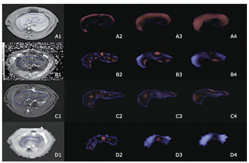
Figure 1.
Table 1
Texture parameters with significant differences between F0 and F1-4 and well as F0-2 and F3-4 comparisons on pre- and post- (60 min delay) contrast T1 mapping images"
| Comparison | Sequence | Texture parameter | SSF | Uvalue | Pvalue |
|---|---|---|---|---|---|
| F0 vs. F1-4 | Pre-contrast T1 mapping | Mean | 4 | 114.0 | 0.004 |
| 5 | 113.0 | 0.006 | |||
| 6 | 109.0 | 0.013 | |||
| Entropy | 0 | 108.0 | 0.015 | ||
| 2 | 107.5 | 0.015 | |||
| 3 | 108.0 | 0.015 | |||
| 4 | 114.0 | 0.013 | |||
| 5 | 113.0 | 0.015 | |||
| 6 | 109.0 | 0.018 | |||
| Post-contrast T1 mapping (20 min) | Mean | 6 | 21.0 | 0.039 | |
| Post-contrast T1 mapping (60 min) | Kurtosis | 6 | 76.5 | 0.025 | |
| F0-2 vs. F3-4 | Pre-contrast T1 mapping | Skewness | 4 | 103.0 | 0.036 |
Table 1
Table 2
Texture parameters with significant differences between F0 and F1-4 as well as F0-2 and F3-4 on pre- and post-contrast T1W images"
| Comparison | Sequence | Texture parameter | SSF | U value | P value |
|---|---|---|---|---|---|
| F0 vs. F1-4 | Pre-contrast T1W | Mean | 2 | 39.0 | 0.045 |
| 3 | 35.0 | 0.025 | |||
| 4 | 37.0 | 0.034 | |||
| MPP | 3 | 34.0 | 0.021 | ||
| 4 | 33.0 | 0.018 | |||
| Post-contrast T1W (20 min) | SD | 0 | 98.0 | 0.013 | |
| Entropy | 0 | 94.5 | 0.023 | ||
| Kurtosis | 2 | 98.5 | 0.011 | ||
| Post-contrast T1W (60 min) | SD | 0 | 40.0 | 0.018 | |
| Entropy | 0 | 39.5 | 0.028 | ||
| MPP | 3 | 6.0 | 0.040 | ||
| 4 | 6.0 | 0.040 | |||
| Skewness | 3 | 41.0 | 0.010 | ||
| 4 | 40.0 | 0.018 | |||
| F0-2 vs. F3-4 | Pre-contrast T1W | Skewness | 0 | 122.0 | 0.041 |
| Post-contrast T1W (20 min) | SD | 0 | 112.0 | 0.004 | |
| Entropy | 0 | 101.5 | 0.027 | ||
| Kurtosis | 2 | 103.0 | 0.023 | ||
| Post-contrast T1W (60 min) | SD | 0 | 52.0 | 0.004 | |
| Entropy | 0 | 51.5 | 0.004 |
Table 2
Table 3
Receiver operating characteristic (ROC) analysis for texture parameters with significant differences of F0 vs. F1-4 and F0-2 vs. F3-4 comparisons on pre- and post-contrast T1 mapping images"
| Sequence | Comparison | Texture parameter | SSF | AUC | 95%CI | Pvalue | Threshold | Sensitivity | Specificity |
|---|---|---|---|---|---|---|---|---|---|
| Pre-contrast T1 mapping | F0 vs. F1-4 | Mean | 4 | 0.857 | 0.711, 1.000 | 0.006 | >-391.855 | 0.684 | 1.000 |
| 5 | 0.850 | 0.701, 0.999 | 0.007 | >-608.24 | 0.789 | 1.000 | |||
| 6 | 0.820 | 0.635, 1.000 | 0.014 | >-745.89 | 0.789 | 0.857 | |||
| Entropy | 0 | 0.812 | 0.638, 0.986 | 0.016 | >5.305 | 0.632 | 1.000 | ||
| 2 | 0.808 | 0.631, 0.985 | 0.018 | >5.655 | 0.632 | 0.857 | |||
| 3 | 0.812 | 0.636, 0.988 | 0.016 | >5.210 | 0.632 | 0.857 | |||
| 4 | 0.816 | 0.641, 0.991 | 0.015 | >5.185 | 0.632 | 0.857 | |||
| 5 | 0.812 | 0.637, 0.987 | 0.016 | >5.225 | 0.632 | 0.857 | |||
| 6 | 0.805 | 0.629, 0.980 | 0.019 | >5.170 | 0.632 | 0.857 | |||
| F0-2 vs.F3-4 | Skewness | 4 | 0.747 | 0.554, 0.939 | 0.037 | >-0.265 | 0.900 | 0.625 | |
| Post-contrast T1 mapping (60 min) | F0 vs. F1-4 | Kurtosis | 6 | 0.797 | 0.592, 1.000 | 0.028 | >-0.390 | 0.833 | 0.750 |
Table 3
Table 4
ROC analysis for texture parameters with significant differences of F0 vs. F1-4 and F0-2 vs. F3-4 comparisons on pre- and post-contrast T1W images"
| Sequence | Comparison | Texture parameter | SSF | AUC | 95%CI | Pvalue | Threshold | Sensitivity | Specificity |
|---|---|---|---|---|---|---|---|---|---|
| Pre-contrast T1W | F0 vs. F1-4 | Mean | 2 | 0.745 | 0.556, 0.934 | 0.043 | <131.57 | 0.765 | 0.556 |
| 3 | 0.771 | 0.586, 0.856 | 0.025 | <186.005 | 0.706 | 0.778 | |||
| 4 | 0.758 | 0.561, 0.955 | 0.033 | <255 | 0.647 | 0.556 | |||
| MPP | 3 | 0.778 | 0.592, 0.964 | 0.022 | <227.22 | 0.706 | 0.778 | ||
| 4 | 0.784 | 0.600, 0.969 | 0.019 | <320.61 | 0.765 | 0.667 | |||
| Post-contrast T1W (20 min) | F0 vs. F1-4 | SD | 0 | 0.817 | 0.642, 0.991 | 0.014 | >53.785 | 0.667 | 1.000 |
| Entropy | 0 | 0.788 | 0.599, 0.976 | 0.026 | >5.04 | 0.667 | 0.825 | ||
| Kurtosis | 2 | 0.821 | 0.644, 0.998 | 0.013 | >0.700 | 0.600 | 0.875 | ||
| F0-2 vs. F3-4 | SD | 0 | 0.848 | 0.689, 1.000 | 0.005 | >53.785 | 0.727 | 0.833 | |
| Entropy | 0 | 0.769 | 0,572, 0.966 | 0.029 | >5.04 | 0.727 | 0.750 | ||
| Kurtosis | 2 | 0.780 | 0.582, 0.979 | 0.023 | >0.155 | 0.818 | 0.593 | ||
| Post-contrast TIW (60 min) | F0 vs. F1-4 | SD | 0 | 0.909 | 0.739, 1.000 | 0.019 | >36.375 | 0.909 | 1.000 |
| Entropy | 0 | 0.898 | 0.735, 1.000 | 0.022 | >4.920 | 0.727 | 1.000 | ||
| MPP | 3 | 0.864 | 0.670, 1.000 | 0.037 | <303.140 | 0.909 | 0.750 | ||
| 4 | 0.864 | 0.671, 1.000 | 0.037 | <351.545 | 0.818 | 1.000 | |||
| Skewness | 3 | 0.932 | 0.794, 1.000 | 0.013 | >-0.115 | 0.909 | 1.000 | ||
| 4 | 0.909 | 0.754, 1.000 | 0.019 | >-0.160 | 0.818 | 1.000 | |||
| F0-2 vs. F3-4 | SD | 0 | 0.929 | 0.782, 1.000 | 0.005 | >37.345 | 1.000 | 0.857 | |
| Entropy | 0 | 0.920 | 0.781, 1.000 | 0.007 | >4.920 | 0.875 | 0.857 | ||
| 3 | 0.804 | 0.569, 1.000 | 0.049 | >6.050 | 0.750 | 0.857 | |||
| 4 | 0.804 | 0.573, 1.000 | 0.049 | >6.095 | 0.625 | 0.857 |
Table 4
| 1. |
Afdhal NH, Nunes D . Evaluation of liver fibrosis: a concise review. Am J Gastroenterol 2004; 99(6):1160-74. doi: 10.1111/j.1572-0241.2004.30110.x.
doi: 10.1111/j.1572-0241.2004.30110.x pmid: 15180741 |
| 2. |
Nguyen D, Talwalkar JA . Noninvasive assessment of liver fibrosis. Hepatology 2011; 53(6):2107-10. doi: 10.1002/hep.24401.
doi: 10.1002/hep.24013 pmid: 21254180 |
| 3. |
Lubner MG, Smith AD, Sandrasegaran K , et al. CT texture analysis: definitions, applications, biologic correlates, and challenges. RadioGraphics 2017; 37(5):1483-503. doi: 10.1148/rg.2017170056.
doi: 10.1148/rg.2017170056 pmid: 28898189 |
| 4. |
Fujimoto K, Tonan T, Azuma S , et al. Evaluation of the mean and entropy of apparent diffusion coefficient values in chronic hepatitis C: correlation with pathologic fibrosis stage and inflammatory activity grade. Radiology 2011; 258(3):739-48. doi: 10.1148/radiol.10100853.
doi: 10.1148/radiol.10100853 pmid: 21248235 |
| 5. |
Barry B, Buch K, Soto JA , et al. Quantifying liver fibrosis through the application of texture analysis to diffusion weighted imaging. Magn Reson Imaging 2014; 32(1):84-90. doi: 10.1016/j.mri.2013.04.006.
doi: 10.1016/j.mri.2013.04.006 pmid: 24239337 |
| 6. |
Bahl G, Cruite I, Wolfson T , et al. Noninvasive classification of hepatic fibrosis based on texture parameters from double contrast-enhanced magnetic resonance images. J Magn Reson Imaging 2012; 36(5):1154-61. doi: 10.1002/jmri.23759.
doi: 10.1002/jmri.23759 pmid: 22851409 |
| 7. |
Jirák D, Dezortová M, Taimr P , et al. Texture analysis of human liver. J Magn Reson Imaging 2002; 15(1):68-74.
doi: 10.1002/jmri.10042 pmid: 11793459 |
| 8. |
Katsube T, Okada M, Kumano S , et al. Estimation of liver function using T1 mapping on Gd-EOB-DTPA-enhanced magnetic resonance imaging. Invest Radiol 2011; 46(4):277-83. doi: 10.1097/RLI.0b013e318200f67d.
doi: 10.1097/RLI.0b013e3182804f84 pmid: 21343827 |
| 9. |
Zhou ZP, Long LL, Huang LJ , et al. Gd-EOB-DTPA-enhanced MRI T1 mapping for assessment of liver function in rabbit fibrosis model: comparison of hepatobiliary phase images obtained at 10 and 20 min. Radiol Med 2017; 122(4):239-47. doi: 10.1007/s11547-016-0719-1.
doi: 10.1007/s11547-016-0719-1 |
| 10. |
Zhou ZP, Long LL, Qiu WJ , et al. Comparison of 10- and 20-min hepatobiliary phase images on Gd-EOB-DTPA-enhanced MRI T1 mapping for liver function assessment in clinic. Abdom Radiol (NY) 2017; 42(9):2272-8. doi: 10.1007/s00261-017-1143-2.
doi: 10.1007/s00261-017-1143-2 pmid: 28396918 |
| 11. |
Ding Y, Rao SX, Zhu T , et al. Liver fibrosis staging using T1 mapping on gadoxetic acid-enhanced MRI compared with DW imaging. Clin Radiol 2015; 70(10):1096-103. doi: 10.1016/j.crad.2015.04.014.
doi: 10.1016/j.crad.2015.04.014 pmid: 26164421 |
| 12. |
Pan S, Wang XQ, Guo QY . Quantitative assessment of hepatic fibrosis in chronic hepatitis B and C: T1 mapping on Gd-EOB-DTPA-enhanced liver magnetic resonance imaging. WJG 2018; 24(18):2024-35. doi: 10.3748/wjg.v24.i18.2024.
doi: 10.3748/wjg.v24.i18.2024 pmid: 29760545 |
| 13. |
Intraobserver and interobserver variations in liver biopsy interpretation in patients with chronic hepatitis C. The French METAVIR Cooperative Study Group. Hepatology 1994; 20(1 Pt 1):15-20.
doi: 10.1002/(ISSN)1527-3350 |
| 14. |
Ganeshan B, Miles KA . Quantifying tumour heterogeneity with CT. Cancer Imaging 2013; 13(1):140-9. doi: 10.1102/1470-7330.2013.0015.
doi: 10.1102/1470-7330.2013.0015 pmid: 23545171 |
| 15. |
Lubner MG, Malecki K, Kloke J , et al. Texture analysis of the liver at MDCT for assessing hepatic fibrosis. Abdom Radiol (NY) 2017; 42(8):2069-78. doi: 10.1007/s00261-017-1096-5.
doi: 10.1007/s00261-017-1096-5 pmid: 28314916 |
| 16. |
Raman SP, Schroeder JL, Huang P , et al. Preliminary data using computed tomography texture analysis for the classification of hypervascular liver lesions: generation of a predictive model on the basis of quantitative spatial frequency measurements—a work in progress. J Comput Assist Tomogr 2015; 39(3):383-95. doi: 10.1097/RCT.0000000000000217.
doi: 10.1097/RCT.0000000000000217 pmid: 25700222 |
| 17. |
Cannella R, Rangaswamy B, Minervini MI , et al. Value of texture analysis on gadoxetic acid-enhanced MRI for differentiating hepatocellular adenoma from focal nodular hyperplasia. AJR Am J Roentgenol 2019; 212(3):538-46. doi: 10.2214/AJR.18.20182.
doi: 10.2214/AJR.18.20182 |
| 18. |
Mulé S, Thiefin G, Costentin C , et al. Advanced hepatocellular carcinoma: pretreatment contrast-enhanced CT texture parameters as predictive biomarkers of survival in patients treated with sorafenib. Radiology 2018; 288(2):445-55. doi: 10.1148/radiol.2018171320.
doi: 10.1148/radiol.2018171320 |
| 19. |
Chen S, Zhu Y, Liu Z , et al. Texture analysis of baseline multiphasic hepatic computed tomography images for the prognosis of single hepatocellular carcinoma after hepatectomy: a retrospective pilot study. Eur J Radiol 2017; 90:198-204. doi: 10.1016/j.ejrad.2017.02.035.
doi: 10.1016/j.ejrad.2017.02.035 pmid: 28583634 |
| 20. |
Canellas R, Mehrkhani F, Patino M , et al. Characterization of portal vein thrombosis (neoplastic versus bland) on CT images using software-based texture analysis and thrombus density (Hounsfield Units). AJR Am J Roentgenol 2016; 207(5):W81-7. doi: 10.2214/AJR.15.15928.
doi: 10.2214/AJR.15.15928 pmid: 27490095 |
| 21. |
Sadot E, Simpson AL, Do RKG , et al. Cholangiocarcinoma: correlation between molecular profiling and imaging phenotypes. PLoS One 2015; 10(7):e0132953. doi: 10.1371/journal.pone.0132953.
doi: 10.1371/journal.pone.0132953 pmid: 26207380 |
| 22. |
Daginawala N, Li B, Buch K , et al. Using texture analyses of contrast enhanced CT to assess hepatic fibrosis. Eur J Radiol 2016; 85(3):511-7. doi: 10.1016/j.ejrad.2015.12.009.
doi: 10.1016/j.ejrad.2015.12.009 pmid: 26860661 |
| 23. |
Romero-Gómez M, Gómez-González E, Madrazo A , et al. Optical analysis of computed tomography images of the liver predicts fibrosis stage and distribution in chronic hepatitis C. Hepatology 2008; 47(3):810-6. doi: 10.1002/hep.22112.
doi: 10.1002/hep.22112 pmid: 18098299 |
| 24. |
Zhang X, Gao X, Liu BJ , et al. Effective staging of fibrosis by the selected texture features of liver: which one is better, CT or MR imaging? Comput Med Imaging Graph 2015; 46 Pt 2: 227-36. doi: 10.1016/j.compmedimag.2015.09.003.
doi: 10.1016/j.compmedimag.2015.09.003 pmid: 26455963 |
| 25. | Jirák D, Dezortová M, Taimr P , et al. Texture analysis of human liver. J Magn Reson Imaging 2002; 15(1):68-74. |
| 26. |
Lubner MG, Stabo N, Lubner SJ , et al. CT textural analysis of hepatic metastatic colorectal cancer: pre-treatment tumor heterogeneity correlates with pathology and clinical outcomes. Abdom Imaging 2015; 40(7):2331-7. doi: 10.1007/s00261-015-0438-4.
doi: 10.1007/s00261-015-0438-4 pmid: 25968046 |
| 27. |
Ng F, Kozarski R, Ganeshan B , et al. Assessment of tumor heterogeneity by CT texture analysis: can the largest cross-sectional area be used as an alternative to whole tumor analysis? Eur J Radiol 2013; 82(2):342-8. doi: 10.1016/j.ejrad.2012.10.023.
doi: 10.1016/j.ejrad.2012.10.023 pmid: 23194641 |
| 28. |
Yoon JH, Lee JM, Paek M , et al. Quantitative assessment of hepatic function: modified look-locker inversion recovery (MOLLI) sequence for T1 mapping on Gd-EOB-DTPA-enhanced liver MR imaging. Eur Radiol 2016; 26(6):1775-82. doi: 10.1007/s00330-015-3994-7.
doi: 10.1007/s00330-015-3994-7 pmid: 26373756 |
| 29. |
Yoon JH, Lee JM, Kang H-J , et al. Quantitative assessment of liver function by using gadoxetic acid-enhanced MRI: hepatocyte uptake ratio. Radiology 2019; 290(1):125-33. doi: 10.1148/radiol.2018180753.
doi: 10.1148/radiol.2018180753 |
| Viewed | ||||||
|
Full text |
|
|||||
|
Abstract |
|
|||||
|