Chinese Medical Sciences Journal ›› 2021, Vol. 36 ›› Issue (4): 323-332.doi: 10.24920/004007
• Reviews • Previous Articles Next Articles
Substitution for In Vitro and In Vivo Tests: Computational Models from Cell Attachment to Tissue Regeneration
Hao Huang1, 4, Chaozong Liu2, Teng Yi3, Maryam Tamaddon2, Shanshan Yuan5, Zhenyun Shi4, *( ), Ziyu Liu1, 2, 3, *()
- 1School of Engineering Medicine, Beihang University, 100191 Beijing, China
2Division of Surgery & Interventional Science, University College London, Royal National Orthopaedic Hospital, Stanmore HA7 4LP, London, UK
3National Institude of Defense Science and Technology, Beijing 100071, China
4School of Mechanical Engineering and Automation, Beihang University, 100191 Beijing, China
5Department of Cardiology, Qingdao Municipal Hospital, Qingdao, Shandong 266071, China
-
Received:2021-09-27Published:2021-12-31Online:2021-12-21 -
Contact:Zhenyun Shi,Ziyu Liu E-mail:shichong1983623@hotmail.com;liu_ziyu@buaa.edu.cn
Cite this article
Hao Huang, Chaozong Liu, Teng Yi, Maryam Tamaddon, Shanshan Yuan, Zhenyun Shi, Ziyu Liu. Substitution for In Vitro and In Vivo Tests: Computational Models from Cell Attachment to Tissue Regeneration[J].Chinese Medical Sciences Journal, 2021, 36(4): 323-332.
share this article
Add to citation manager EndNote|Reference Manager|ProCite|BibTeX|RefWorks
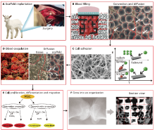
Figure 1.
Schematic diagram of the whole process of bone tissue engineering. A. Bone tissue engineering scaffold implantation;[9] B. Blood filling after implantation;[9, 10] C. Cells adhesion;[11, 12] D. Blood coagulation;[10] E. Cell proliferation, differentiation and migration;[13] F. Formation of an organization.[9]"
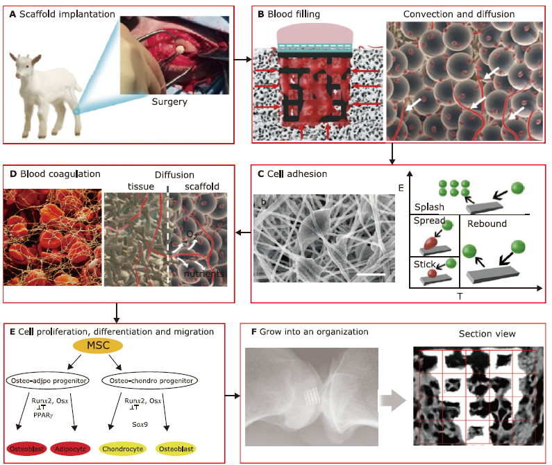
Figure 1.
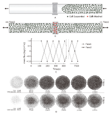
Figure 2.
Dynamic conditions and results of Olivares et al.’s research.[6] A. Suspension process at a constant velocity of 1 mm/s, representation of area covered by the cells after last injection; the cells do not touch the scaffold wall. B. Dispersion of cells after oscillating fluid flow. C. Graphical representation of initial injection conditions and cycles repeating during 2 h of experimentation. Maximum mass flux corresponds to maximum fluid normal velocity of un=1 mm/s. D. Superior view of cells attached in the type I (top) and type G (bottom) scaffolds. The evolution of increment of cell numbers deposited in each time selected."

Figure 2.

Figure 3.
Stanton-Rutland model proposed by Liu et al.[12] A. Impinge regimes definition. B. Cells growth on porous titanium matrix was examined by confocal microscopy, and the confocal image was further processed to determine the cells distribution. C. Predicted cell distribution during cell seeding process. From left to right represent the cells distribution at second one, two, three, four, five and six, respectively."
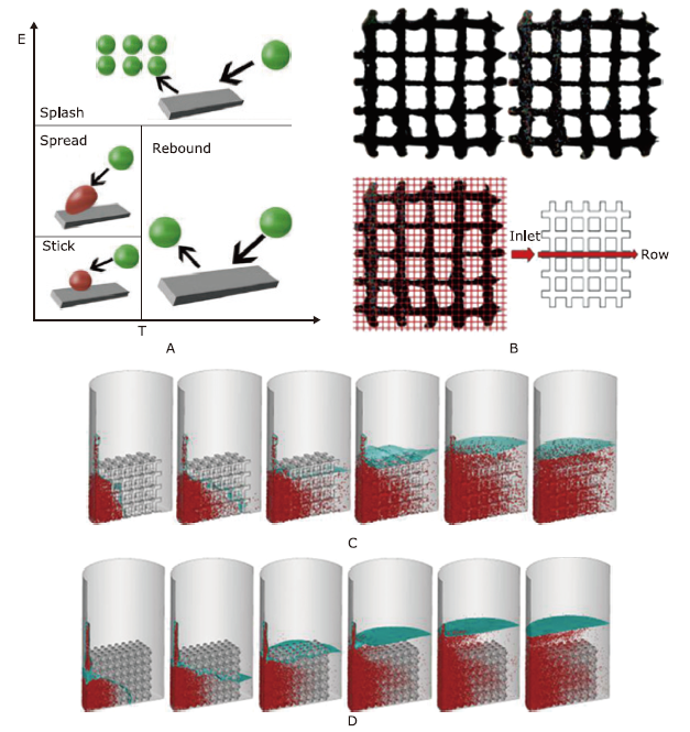
Figure 3.

Figure 4.
The research of Pérez et al.[34] A. Possible position that daughter cells can occupy after mitosis. The distance between sites is only schematic; adjacent sites in the algorithm are considered to be exactly the diameter of a mesenchymal stem cell.[36] B. Possible position that daughter cells can occupy after mitosis with anisotropic proliferation direction (preferred direction is given by the arrow)."
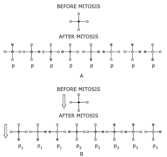
Figure 4.
Figure 5.
The research of Byrne et al.[8] A. Finite element model of a 50% porous scaffold with regular porosity (green). Only one-eighth needs to be modelled because of symmetry (yellow box). The cavity is initially occupied by granulation tissue (in red). B. Lattice generated for each granulation element to model cellular activity. C. Increasing lattice points to account for dissolution of scaffold material."

Figure 5.
| 1. |
Kang H, Zeng Y, Varghese S. Functionally graded multilayer scaffolds for in vivo osteochondral tissue engineering. Acta Biomater 2018; 78:365-77. doi: 10.1016/j.actbio.2018.07.039.
doi: 10.1016/j.actbio.2018.07.039 |
| 2. |
Nooeaid P, Salih V, Beier JP, et al. Osteochondral tissue engineering: scaffolds, stem cells and applications. J Cell Mol Med 2012; 16(10):2247-70. doi: 10.1111/j.1582-4934.2012.01571.x.
doi: 10.1111/j.1582-4934.2012.01571.x |
| 3. |
Yang PJ, Temenoff JS. Engineering orthopedic tissue interfaces. Tissue Eng Part B Rev 2009; 15(2):127-41. doi: 10.1089/ten.teb.2008.0371.
doi: 10.1089/ten.teb.2008.0371 |
| 4. |
Hunziker EB. Articular cartilage repair: basic science and clinical progress. A review of the current status and prospects. Osteoarthritis Cartilage 2002; 10(6):432-63. doi: 10.1053/joca.2002.0801.
doi: 10.1053/joca.2002.0801 |
| 5. |
Xu S, Du P, Xie Y, et al. Cell distribution in a scaffold with random architectures under the influence of fluid dynamics. J Biomater Appl 2008; 23(3):229-45. doi: 10.1177/0885328207086322.
doi: 10.1177/0885328207086322 |
| 6. |
Olivares AL, Lacroix D. Simulation of cell seeding within a three-dimensional porous scaffold: a fluid-particle analysis. Tissue Eng Part C Methods 2012; 18(8):624-31. doi: 10.1089/ten.TEC.2011.0660.
doi: 10.1089/ten.TEC.2011.0660 |
| 7. |
Prendergast PJ, Huiskes R, Søballe K. ESB Research Award 1996. Biophysical stimuli on cells during tissue differentiation at implant interfaces. J Biomech 1997; 30(6):539-48. doi: 10.1016/s0021-9290(96)00140-6.
doi: 10.1016/s0021-9290(96)00140-6 pmid: 9165386 |
| 8. |
Byrne DP, Lacroix D, Planell JA, et al. Simulation of tissue differentiation in a scaffold as a function of porosity, Young’s modulus and dissolution rate: application of mechanobiological models in tissue engineering. Biomaterials 2007; 28(36):5544-54. doi: 10.1016/j.biomaterials.2007.09.003.
doi: 10.1016/j.biomaterials.2007.09.003 |
| 9. |
Liu Z, Tamaddon M, Chen SM, et al. Determination of an initial stage of the bone tissue ingrowth into titanium matrix by cell adhesion model. Front Bioeng Biotechnol 2021; 9:736063. doi: 10.3389/fbioe.2021.736063.
doi: 10.3389/fbioe.2021.736063 |
| 10. |
Liu J, Chen G, Xu H, et al. Pre-vascularization in fibrin Gel/PLGA microsphere scaffolds designed for bone regeneration. NPG Asia Materials 2018; 10(8):827-39. doi: 10.1038/s41427-018-0076-8.
doi: 10.1038/s41427-018-0076-8 |
| 11. |
Daley WP, Peters SB, Larsen M. Extracellular matrix dynamics in development and regenerative medicine. J Cell Sci 2008; 121(Pt 3):255-64. doi: 10.1242/jcs.006064.
doi: 10.1242/jcs.006064 |
| 12. |
Liu Z, Tamaddon M, Gu Y, et al. Cell seeding process experiment and simulation on three-dimensional polyhedron and cross-link design scaffolds. Front Bioeng Biotechnol 2020; 8:104. doi: 10.3389/fbioe.2020.00104.
doi: 10.3389/fbioe.2020.00104 |
| 13. |
Ali F, Taresh S, Al-Nuzaily M, et al. Stem cells differentiation and probing their therapeutic applications in hematological disorders: a critical review. Eur Rev Med Pharmacol Sci 2016; 20(20):4390-400. doi: 10.1155/2016/7653489.
doi: 10.1155/2016/7653489 |
| 14. |
Santoro R, Olivares AL, Brans G, et al. Bioreactor based engineering of large-scale human cartilage grafts for joint resurfacing. Biomaterials 2010; 31(34):8946-52. doi: 10.1016/j.biomaterials.2010.08.009.
doi: 10.1016/j.biomaterials.2010.08.009 |
| 15. | Wendt D, Stroebel S, Jakob M, et al. Uniform tissues engineered by seeding and culturing cells in 3D scaffolds under perfusion at defined oxygen tensions. Biorheology 2006; 43(3, 4):481-8. |
| 16. |
Cherry EM, JK Eaton. Shear thinning effects on blood flow in straight and curved tubes. Phys FLUIDS 2013; 25:0733104. doi: 10.1063/1.4816369.
doi: 10.1063/1.4816369 |
| 17. |
Reymond P, Perren F, Lazeyras F, et al. Patient-specific mean pressure drop in the systemic arterial tree, a comparison between 1-D and 3-D models. J Biomech 2012; 45(15):2499-505. doi: 10.1016/j.jbiomech.2012.07.020.
doi: 10.1016/j.jbiomech.2012.07.020 |
| 18. |
Truscello S, Kerckhofs G, Van Bael S, et al. Prediction of permeability of regular scaffolds for skeletal tissue engineering: a combined computational and experimental study. Acta Biomater 2012; 8(4):1648-58. doi: 10.1016/j.actbio.2011.12.021.
doi: 10.1016/j.actbio.2011.12.021 pmid: 22210520 |
| 19. |
Xiang J, Tremmel M, Kolega J, et al. Newtonian viscosity model could overestimate wall shear stress in intracranial aneurysm domes and underestimate rupture risk. J Neurointerv Surg 2012; 4(5):351-7. doi: 10.1136/neurintsurg-2011-010089.
doi: 10.1136/neurintsurg-2011-010089 |
| 20. |
Jiang Y, Zhang J, Zhao W. Effects of the inlet conditions and blood models on accurate prediction of hemodynamics in the stented coronary arteries. AIP Adv 2015; 5(5):057109. doi: 10.1063/1.4919937.
doi: 10.1063/1.4919937 |
| 21. |
Koch MA, Vrij EJ, Engel E, et al. Perfusion cell seeding on large porous PLA/calcium phosphate composite scaffolds in a perfusion bioreactor system under varying perfusion parameters. J Biomed Mater Res A 2010; 95(4):1011-8. doi: 10.1002/jbm.a.32927.
doi: 10.1002/jbm.a.32927 pmid: 20872752 |
| 22. |
Wendt D, Marsano A, Jakob M, et al. Oscillating perfusion of cell suspensions through three-dimensional scaffolds enhances cell seeding efficiency and uniformity. Biotechnol Bioeng 2003; 84(2):205-14. doi: 10.1002/bit.
doi: 10.1002/bit pmid: 12966577 |
| 23. |
Stanton DW, CJ Rutland. Modeling fuel film formation and wall interaction in diesel engines. SAE Trans 1996; 808-24. doi: 10.4271/960628.
doi: 10.4271/960628 |
| 24. | O’rourke PJ, Amsden AA. A particle numerical model for wall film dynamics in port-injected engines. SAE Technical Paper Series. Detroit: SAE; 1996. p.2000-13. |
| 25. |
O’Rourke PJ, Amsden AA. A spray/wall interaction submodel for the KIVA-3 wall film model. SAE Technical Paper Series. Detroit: SAE; 2000. p.281-98. doi: 10.4271/2000-01-0271.
doi: 10.4271/2000-01-0271 |
| 26. |
Karande TS, Ong JL, Agrawal CM. Diffusion in musculoskeletal tissue engineering scaffolds: design issues related to porosity, permeability, architecture, and nutrient mixing. Ann Biomed Eng 2004; 32(12):1728-43. doi: 10.1007/s10439-004-7825-2.
doi: 10.1007/s10439-004-7825-2 pmid: 15675684 |
| 27. |
Makhaniok A, Haranava Y, Goranov V, et al. In silico prediction of the cell proliferation in porous scaffold using model of effective pore. Biosystems 2013; 114(3):227-37. doi: 10.1016/j.biosystems.2013. 10.001.
doi: 10.1016/j.biosystems.2013.10.001 pmid: 24141144 |
| 28. |
Dunn JC, Chan WY, Cristini V, et al. Analysis of cell growth in three-dimensional scaffolds. Tissue Eng 2006; 12(4):705-16. doi: 10.1089/ten.2006.12.705.
doi: 10.1089/ten.2006.12.705 |
| 29. |
Rouwkema J, Koopman B, Blitterswijk C, et al. Supply of nutrients to cells in engineered tissues. Biotechnol Genet Eng Rev 2010; 26:163-78. doi: 10.5661/bger-26-163.
doi: 10.5661/bger-26-163 pmid: 21415880 |
| 30. |
Reynolds M, McCann SR. Human marrow stromal cells in short-term semi-solid bone marrow culture in aplastic anaemia. Scand J Haematol 1985; 34(2):101-10. doi: 10.1111/j.1600-0609.1985.tb02241.x.
doi: 10.1111/j.1600-0609.1985.tb02241.x pmid: 3975567 |
| 31. |
Winterton RHS. Thermal design of nuclear reactors. Elsevier; 1981. Available from: https://doi.org/10.1016/B978-0-08-024215-6.50003-6.
doi: https://doi.org/10.1016/B978-0-08-024215-6.50003-6 |
| 32. |
Botchwey EA, Dupree MA, Pollack SR, et al. Tissue engineered bone: measurement of nutrient transport in three-dimensional matrices. J Biomed Mater Res A 2003; 67(1):357-67. doi: 10.1002/jbm.a.10111.
doi: 10.1002/jbm.a.10111 pmid: 14517896 |
| 33. |
Mofrad AZ, Mashayekhan S, Bastani D. Simulation of the effects of oxygen carriers and scaffold geometry on oxygen distribution and cell growth in a channeled scaffold for engineering myocardium. Math Biosci 2017; 294:160-71. doi: 10.1016/j.mbs.2017.09.003.
doi: 10.1016/j.mbs.2017.09.003 |
| 34. |
Pérez MA, Prendergast PJ. Random-walk models of cell dispersal included in mechanobiological simulations of tissue differentiation. J Biomech 2007; 40(10):2244-53. doi: 10.1016/j.jbiomech.2006.10.020.
doi: 10.1016/j.jbiomech.2006.10.020 pmid: 17173925 |
| 35. |
Søballe K, Hansen ES, B-Rasmussen H, et al. Tissue ingrowth into titanium and hydroxyapatite-coated implants during stable and unstable mechanical conditions. J Orthop Res 1992; 10(2):285-99. doi: 10.1002/jor.1100100216.
doi: 10.1002/jor.1100100216 pmid: 1311039 |
| 36. |
Lanza R, Gearhart J, Hogan B, et al. Essentials of stem cell biology. Elsevier; 2009. Available from: https://doi.org/10.1016/C2009-0-00078-6.
doi: https://doi.org/10.1016/C2009-0-00078-6 |
| [1] | Zong-ming Wan, Lu Liu, Jian-yu Li, Rui-xin Li, Yong Guo, Hao Li, Jian-ming Zhang, Xi-zheng Zhang. Mechanical Stimulus Inhibits the Growth of a Bone Tissue Model Cultured In Vitro [J]. Chinese Medical Sciences Journal, 2013, 28(4): 218-224. |
| Viewed | ||||||
|
Full text |
|
|||||
|
Abstract |
|
|||||
|