延髓发病型及脊髓发病型肌萎缩侧索硬化症脑部磁共振结构特征变化
Gray Matter Volume Changes over the Whole Brain in the Bulbar- and Spinal-onset Amyotrophic Lateral Sclerosis: a Voxel-based Morphometry Study
延髓发病型及脊髓发病型肌萎缩侧索硬化症脑部磁共振结构特征变化 |
| 陈志晔,刘梦琦,马林 |
|
Gray Matter Volume Changes over the Whole Brain in the Bulbar- and Spinal-onset Amyotrophic Lateral Sclerosis: a Voxel-based Morphometry Study |
| Chen Zhiye,Liu Mengqi,Ma Lin |
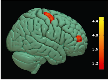
| Figure 1. Brain region with decreased gray matter (GM) volume in ALS group compared with NC group. Colored areas represent the involved brain areas: right precentral gyrus and middle frontal gyrus respectively. The value of the color bar represents T value. |
|
|