影像学诊断1例III型胆总管囊肿
Imaging Diagnosis of Type Ⅲ Choledochal Cyst: A Case Report
影像学诊断1例III型胆总管囊肿 |
| 李平,朱亮,王萱,薛华丹,吴晰,金征宇 |
|
Imaging Diagnosis of Type Ⅲ Choledochal Cyst: A Case Report |
| Li Ping,Zhu Liang,Wang Xuan,Xue Huadan,Wu Xin,Jin Zhengyu |
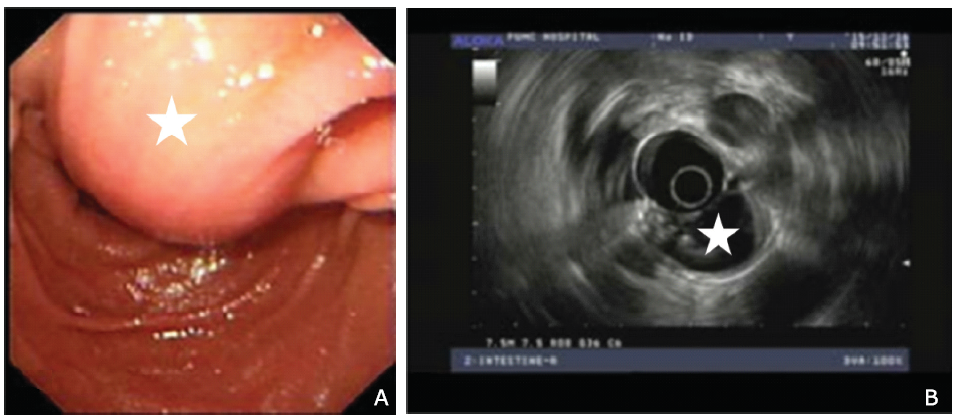
| Figure 1. Endoscopy (A) revealed a parenteral cystic lesion (star) in the descending duodenum. Endoscopic ultrasonography (B) showed a cystic lesion in the duodenal lumen with many stones (star). |
|
|