影像学诊断1例III型胆总管囊肿
Imaging Diagnosis of Type Ⅲ Choledochal Cyst: A Case Report
影像学诊断1例III型胆总管囊肿 |
| 李平,朱亮,王萱,薛华丹,吴晰,金征宇 |
|
Imaging Diagnosis of Type Ⅲ Choledochal Cyst: A Case Report |
| Li Ping,Zhu Liang,Wang Xuan,Xue Huadan,Wu Xin,Jin Zhengyu |
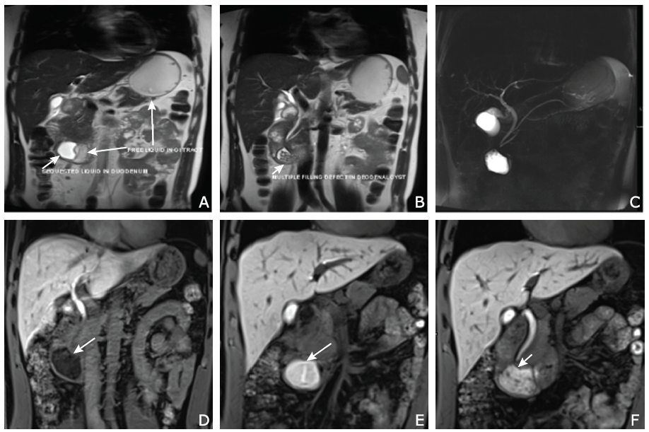
| Figure 4. Magnetic resonance cholangiopancreatography (MRCP) (A-C) and dynamic contrast enhancement (DCE)-MRI (D-F) images. A. Pineapple juice can reduce the pre-existing gastrointestinal tract liquid (long arrow) in T2 signal and distinguish bowel water from liquid in the cystic lesion (short arrow) on T2WI. B. MRCP showing multiple filling defects in the cystic cavity (arrow). C. 2D MRCP. D-F. DCE-MRI revealing the process of contrast medium excreted into the cystic lesion during discharge phase (arrows) and clearly displaying a slender tube (arrow, F) connecting the lower common bile duct with the cystic lesion. |
|
|