纵向测量大鼠非动脉炎性前部缺血性视神经病变模型中后部视乳头血流变化的研究
Longitudinal Measurement of Hemodynamic Changes within the Posterior Optic Nerve Head in Rodent Nonarteritic Anterior Ischemic Optic Neuropathy
纵向测量大鼠非动脉炎性前部缺血性视神经病变模型中后部视乳头血流变化的研究 |
| 马瑾,陈婷,王一玮,赵潺,李东辉,王萌,干霖洋,钟勇 |
|
Longitudinal Measurement of Hemodynamic Changes within the Posterior Optic Nerve Head in Rodent Nonarteritic Anterior Ischemic Optic Neuropathy |
| Ma Jin,Chen Ting,Wang Yiwei,Zhao Chan,Li Donghui,Wang Meng,Gan Linyang,Zhong Yong |
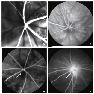
| Figure 2. Representative FFA images of a naive eye and an rNAION eye 3 hours after disease induction. A. normal fluorescein perfusion of the choroidal and retinal vasculature in the early phase of FFA in a naive eye. B. normal fluorescein distribution in the fundus in the late phase of FFA in a naive eye; C. FFA imaging 3 seconds after injection (early phase) of an rNAION eye showed filling defects in the choroid and the ONH (arrow); D. late phase imaging of the same eye showed marked dye leakage (arrow) from the ONH. FFA: fundus fluorescein angiography. |
|
|