纵向测量大鼠非动脉炎性前部缺血性视神经病变模型中后部视乳头血流变化的研究
Longitudinal Measurement of Hemodynamic Changes within the Posterior Optic Nerve Head in Rodent Nonarteritic Anterior Ischemic Optic Neuropathy
纵向测量大鼠非动脉炎性前部缺血性视神经病变模型中后部视乳头血流变化的研究 |
| 马瑾,陈婷,王一玮,赵潺,李东辉,王萌,干霖洋,钟勇 |
|
Longitudinal Measurement of Hemodynamic Changes within the Posterior Optic Nerve Head in Rodent Nonarteritic Anterior Ischemic Optic Neuropathy |
| Ma Jin,Chen Ting,Wang Yiwei,Zhao Chan,Li Donghui,Wang Meng,Gan Linyang,Zhong Yong |
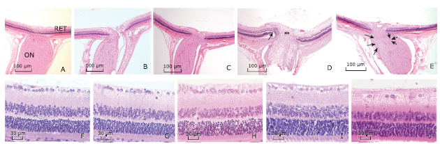
| Figure 3. Representative histologic sections of ONH and retina in naive and rNAION eyes (H&E stain). A. longitudinal sections of the ONH in naive eyes, B. in the laser-only eyes and C. in the RB-only eyes showed normal histology and anatomy of the optic nerve and peripapillary retina. D. On day 1 after rNAION induction, the ONH was edematous with thickened nerve fiber bundles (double asterisks) and peripapillary retinal detachment (arrow). E. On day 90 after induction, there was a reduced number of RGC axons (long arrow) accompanied by gliosis and cellular infiltration (short arrow). F. naive eyes, G. laser-only eyes, H. RB-only eyes, and I. eyes 1 day after rNAION induction. The RGCs in peripapillary retina were closely packed in a single layer with normal density. J. On day 90 after rNAION induction, there was an obvious reduction in density of the RGCs in retina, while the cell densities in the INL and ONL remained mainly unchanged. (magnifying power: A-E, 50×; F-J, 200×). RB: rose bengal; RGC: retinal ganglion cell; INL: inner nuclear layer; ONL: outer nuclear layer. |
|
|