Chinese Medical Sciences Journal ›› 2023, Vol. 38 ›› Issue (2): 138-146.doi: 10.24920/004159
• Review • Previous Articles Next Articles
Central Granular Cell Odontogenic Tumor: A Literature Review of Cases Reported in the Last 71 Years with a New Case Report
Fatemeh Mashhadiabbas1, Sanaz GholamiToghchi1, Roohollah Safarpour2( )
)
- 1Department of Oral and Maxillofacial Pathology, School of Dentistry, Shahid Beheshti University of Medical Sciences, Tehran 1983969413, Iran
2Department of Oral and Maxillofacial Pathology, School of Dentistry, Lorestan University of Medical Sciences, Khorramabad 6813833946, Iran
-
Received:2022-08-19Accepted:2023-03-14Published:2023-06-30Online:2023-04-14 -
Contact:*98-917-7737642, 98-663-3207826, E-mail:Safarpoor.r@lums.ac.ir ,roohollah.safarpour@gmail.com
Cite this article
Fatemeh Mashhadiabbas, Sanaz GholamiToghchi, Roohollah Safarpour. Central Granular Cell Odontogenic Tumor: A Literature Review of Cases Reported in the Last 71 Years with a New Case Report[J].Chinese Medical Sciences Journal, 2023, 38(2): 138-146.
share this article
Add to citation manager EndNote|Reference Manager|ProCite|BibTeX|RefWorks

Figure 1.
A photograph showing a swelling in the anterior region of the right maxilla (arrows) of a 39-year-old white female with CGCOT. CGCOT: central granular cell odontogenic tumor."

Figure 1.

Figure 2.
Cone beam computed tomography images of a 39-year-old white female with CGCOT. A well-defined corticated unilocular radiolucent lesion (arrows) extending from maxillary right central incisor to the right first premolar is noted."
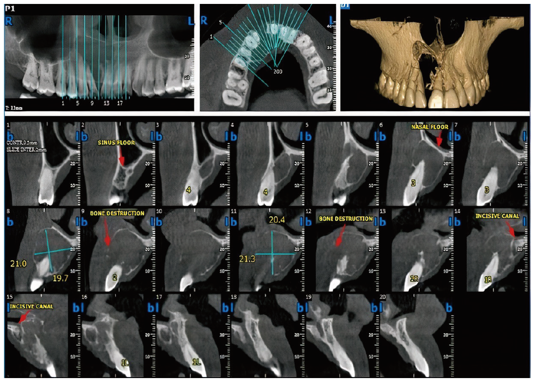
Figure 2.

Figure 3.
Histopathological results of a 39-year-old white female with CGCOT. HE staining (A) Sheets and lobules of eosinophilic granular cells are intermixed with cords and strands of odontogenic epithelium. (B) Large granular cells with eccentric vesicular nuclei and vacuolated odontogenic epithelium (arrow)."
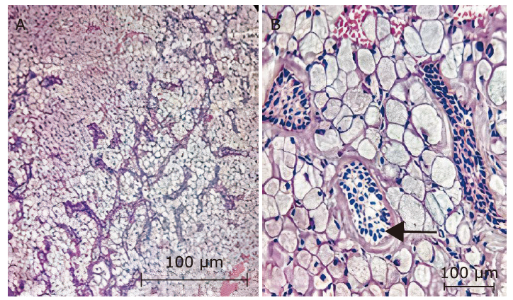
Figure 3.

Figure 4.
Immunohistochemical results of a 39-year-old white female with CGCOT. EnVision staining (A) Granular cells are positive for CD68, and odontogenic epithelium is negative for CD68. (B) Granular cells are positive for vimentin, whereas odontogenic epithelium is negative for vimentin. (C) Granular cells are negative for S-100."

Figure 4.
Table 1.
Demographic and clinical findings of 51 reported cases with CGCOT in the literatures"
| References | Year of publication | Age (yrs) | Gender | Location | Radiographic features | Treatment | Follow-up |
|---|---|---|---|---|---|---|---|
| Werthemann[ | 1950 | 39 | Male | Left mandibular premolar/molar | NS | NS | NS |
| Couch et al.[ | 1962 | 55 | Femle | Left mandibular second molar | Radiolucent lesion with loculated borders | Conservative removal of the lesion with tooth extraction | NR 8 months |
| Couch et al.[ | 1962 | 59 | Female | Left mandibular canine | Loculated radiolucency with focal densities | Removal of tumor | NR 27 months |
| Waldron et al.[ | 1963 | 60 | Female | Left mandibular canine | 2.0 cm radiolucent lesion | Removal of tumor | NR 29 months |
| Waldron et al.[ | 1963 | 53 | Female | Left mandibular molar | 2.0-3.0 cm cystic radiolucency, displacment of teeth | Removal of the mass with tooth extraction | NR 3 months |
| Gorlin et al.[ | 1970 | 50 | Female | Mandibular molar region | NS | Curettage | NS |
| Dalforno et al.[ | 1970 | 57 | NS | Left mandibular molar | NS | Curettage | NR 6 months |
| White et al.[ | 1978 | 50 | Female | Mandibular canine area | Radiolucency | Curettage | NR 6 months |
| White et al.[ | 1978 | 50 | Female | Mandibular posterior area | Radiolucency | Curettage | NR 7 years |
| White et al.[ | 1978 | 55 | Female | Maxillary premolar | Radiolucency | Surgical excision | NR 3 years |
| White et al.[ | 1978 | 65 | Female | Mandibular premolar/molar | Radiolucency | Surgical excision | NR 2 years |
| Regezi et al.[ | 1978 | 29 | Female | Maxilla | NS | NS | NS |
| Regezi et al.[ | 1978 | 16 | Male | Mandible | NS | NS | NS |
| Vincent et al.[ | 1987 | 51 | Female | Right mandibular premolar/molar | 4-2 cm radiolucency with sclerotic border | Conservative removal of the mass | NS |
| Vincent et al.[ | 1987 | 27 | Male | Right mandibular second premolar/first molar | 1.5 cm unicystic radiolucency with sclerotic borders | Surgical excision | NR 24 months |
| Shiro et al.[ | 1989 | 45 | Female | Left mandibular premolars | 0.7-0.4 cm unicystic radiolucency | Surgical excision | NR 4 years |
| Mirchandani et al.[ | 1989 | 33 | Female | Mandible | Radiolucency | NS | NS |
| Ruhl et al.[ | 1989 | 22 | Male | Left maxillary first and second molars | 4.5 cm with slight displacement of teeth | Complete resection | NS |
| Chen[ | 1991 | 50 | Female | Right mandibular canine | 1.0-0.8 cm radiolucency | NS | NS |
| Chen[ | 1991 | 45 | Female | Left mandibular premolar/molar | 5.0-3.0 cm radiolucency | NS | NS |
| Chen[ | 1991 | 64 | Female | Left mandibular canine/premolar | 3.0-2.0 cm radiolucency | NS | NS |
| Chen[ | 1991 | 77 | Female | Left mandibular premolars | 0.5-0.5 cm radiolucency | NS | NS |
| Yih et al.[ | 1995 | 66 | Female | Left mandibular second premolar | 0.5-0.5 cm unilocular radiolucency | Curettage | NR 6 months |
| Gesek et al.[ | 1995 | 62 | Female | Left mandibular second premolar | Multilocular, well-circumscribed radiolucency | Curettage | NR 12 months |
| Machado de Sousa et al.[ | 1998 | 19 | Female | Right maxillary premolar/molar | Well-delineated multilocular radiolucency | Surgical excision | NR 24 months |
| Machado de Sousa et al.[ | 1998 | 25 | Male | Right maxillary premolar/molar | 8.0 cm radiopaque lesion | Surgical excision | NR 120 months |
| Ardekian et al.[ | 1998 | 63 | Male | Right maxillary premolar/molar | Well-defined radiolucency with sclerotic border | Curettage Teeth extraction | NR 48 months |
| Matsumoto et al.[ | 2000 | 24 | Male | Left mandibular premolars | Well-demarcated radiolucent lesion | Enucleation with teeth extraction | NR 1.5 years |
| Brannon et al.[ | 2002 | 36 | Female | Mandibular canine/ premolar | NS | NS | NS |
| Brannon et al.[ | 2002 | 50 | Female | Jaw | NS | NS | NS |
| Brannon et al.[ | 2002 | 32 | Female | Mandibular canine/ premolar | Multilocular radiolucency with sclerotic border | Teeth extraction with surgical excision | NR 180 months |
| Brannon et al.[ | 2002 | 19 | Female | Left maxillary first premolar/first molar | Unicystic radiolucency enveloping roots of second premolar | Curettage | Recurrence 156 months |
| Brannon et al.[ | 2002 | 48 | Male | Right side of maxilla | NS | NS | NS |
| Calvo et al.[ | 2002 | 61 | Male | Anterior region of maxilla | Radiolucency with resorption of anterior teeth | NS | NS |
| Meer et al.[ | 2004 | 65 | Female | Left mandibular first premolar/second molar | Irregular radiolucency from first premolar to second molar | Surgical excision | NR 12 months |
| Reichart et al.[ | 2006 | 46 | Female | Right mandibular premolar/molar | Multilocular radiolucent lesion | Surgical excision with reconstruction | NR 2 yrs. |
| Gomes et al.[ | 2006 | 20 | Female | Left mandibular premolars/molars | An intra-osseous mixed lesion, 5 cm | Enucleation | NR 7 months |
| Kim et al.[ | 2006 | 33 | Male | Right maxillary premolar/first molar | Well-defined unilocular radiolucency | Enucleation with tooth extraction | NR 23 months |
| Mesquita et al.[ | 2009 | 20 | Female | Left mandibular second premolar/second molar | Well-defined radiolucency with foci of calcifications | Complete resection of tumor | NR 4 years |
| Lotay et al.[ | 2010 | 28 | Female | Right maxillary premolar | 1.5-2.5 cm well-defined mixed lesion | Enucleation and curettage | NS |
| Silva et al.[ | 2012 | 41 | Female | Left side of maxilla | Well-defined mixed lesion | Surgical excision | NR 2 years |
| Sarode et al.[ | 2013 | 25 | Female | Right side of mandible crossing the midline | Well-demarcated multilocular radiolucent lesion | Enucleation and curettage | NR 2 years |
| Cheng et al.[ | 2013 | 52 | Female | Right mandibular premolars | Well-defined mixed lesion | Enucleation | NR 3 months |
| Chiang et al.[ | 2014 | 69 | Male | Ramus of left mandible | Well-demarcated radiolucent lesion | Surgical excision | NR 2 months |
| Anbiaee et al.[ | 2014 | 16 | Female | Left mandibular angle | Multilocular mixed lesion, 3 cm × 5 cm | Surgical resection with mandibular reconstruction | NR 2 years |
| Lee et al.[ | 2014 | 19 | Male | Left mandibular third molar | Enlarged dental follicle | Enucleation with tooth extraction | NS |
| Fletcher et al.[ | 2015 | 19 | Female | Right mandibular second premolar/molars | Unilocular radiolucent lesion | Curettage | NR 24 months |
| Vennamaneni et al.[ | 2016 | 38 | Male | Right mandibular premolars/first molar | Well-defined unilocular radiolucent lesion | Enucleation | NR NS |
| Madan et al.[ | 2016 | 73 | Male | Anterior area of mandible | Multilocular radiolucent lesion | Segmental resection | NR 9 months |
| Atarbashi- Moghadam et al.[ | 2019 | 57 | Female | Left mandibular premolars/first molar | Well defined radiolucent lesion | Enucleation | NR 12 months |
| Koth et al.[ | 2021 | 42 | Femlae | Left maxillary anterior region | Unilocular radiolucency | Surgical removal | NR 16 months |
Table 1.
| 1 |
Werthemann A. Spongiocytic adamantinoma. Oncologia 1950; 3 (4):193-207. doi: 10.1159/000223592.
doi: 10.1159/000223592 |
| 2 |
Couch RD, Morris EE, Vellios F. Granular cell ameloblastic fibroma. Report of 2 cases in adults, with observations of its similarity to congenital epulis. Am J Clin Pathol 1962 ; 37(4):398-404. doi: 10.1093/ajcp/37.4.398.
doi: 10.1093/ajcp/37.4.398 |
| 3 | Dalforno S, Donna A. Soft odontoma (ameloblastic fibroma) with granular cell stroma. Cancro 1970 ; 23(1):61-6. Italian. |
| 4 |
White DK, Chen SY, Hartman KS, et al. Central granular-cell tumor of the jaws (the so-call granular-cell ameloblastic fibroma). Oral Surg Oral Med Oral Pathol 1978 ; 45(3):396-405. doi: 10.1016/0030-4220(78)90525-x.
doi: 10.1016/0030-4220(78)90525-x |
| 5 |
Vincent SD, Hammond HL, Ellis GL, et al. Central granular cell odontogenic fibroma. Oral Surg Oral Med Oral Pathol 1987 ; 63(6):715-21. doi: 10.1016/0030-4220(87)90376-8.
doi: 10.1016/0030-4220(87)90376-8 |
| 6 |
Shiro BC, Jacoway JR, Mirmiran SA, et al. Central odontogenic fibroma, granular cell variant. A case report with S-100 immunohistochemistry and a review of the literature. Oral Surg Oral Med Oral Pathol 1989 ; 67(6):725-30. doi: 10.1016/0030-4220(89)90015-7.
doi: 10.1016/0030-4220(89)90015-7 |
| 7 |
Mirchandani R, Sciubba JJ, Mir R. Granular cell lesions of the jaws and oral cavity: a clinicopathologic, immunohistochemical, and ultrastructural study. J Oral Maxillofac Surg 1989 ; 47(12):1248-55. doi: 10.1016/0278-2391(89)90718-0.
doi: 10.1016/0278-2391(89)90718-0 |
| 8 |
Gesek DJ Jr, Adrian JC, Reid EN. Central granular cell odontogenic tumor: a case report including light microscopy, immunohistochemistry, and literature review. J Oral Maxillofac Surg 1995 ; 53(8):945-9. doi: 10.1016/0278-2391(95)90290-2.
doi: 10.1016/0278-2391(95)90290-2 |
| 9 |
Gomes CC, Naves MD, Pereira MV, et al. Granular cell odontogenic tumor: case report and review of literature. Oral Oncol Extra 2006; 42:277-80. doi: 10.1016/j.ooe.2006.07.001.
doi: 10.1016/j.ooe.2006.07.001 |
| 10 |
Sarode SC, Sarode GS, Vaidya K. Central granular cell odontogenic tumor: a systematic review. J Oral Pathol Med 2014 ; 43(3):167-76. doi: 10.1111/jop.12085.
doi: 10.1111/jop.12085 |
| 11 |
Ardekian L, Manor R, Gaspar R, et al. Central granular cell odontogenic tumor: case report and review of literature. J Oral Maxillofac Surg 1998 ; 56(11):1343-5. doi: 10.1016/s0278-2391(98)90619-x.
doi: 10.1016/s0278-2391(98)90619-x |
| 12 |
Brannon RB, Goode RK, Eversole LR, et al. The central granular cell odontogenic tumor: report of 5 new cases. Oral Surg Oral Med Oral Pathol Oral Radiol Endod 2002 ; 94(5):614-21. doi: 10.1067/moe.2002.126890.
doi: 10.1067/moe.2002.126890 |
| 13 |
Meer S, Altini M, Coleman H, et al. Central granular cell odontogenic tumor: immunohistochemistry and ultrastructure. Am J Otolaryngol 2004 ; 25(1):73-8. doi: 10.1016/j.amjoto.2003.09.002.
doi: 10.1016/j.amjoto.2003.09.002 |
| 14 | Kim JW, Park IS, Byeon GJ, et al. Central granular cell odontogenic tumor (CGCOT): a case report including light microscopy, immunohistochemistry and literature review. J Korean Assoc Oral Maxillofac Surg 2006 ; 32(4):374-9. |
| 15 |
Mesquita AT, Santos CR, Gomez RS, et al. Central granular cell odontogenic tumor: a histopathologic and immunohistochemical study. Ann Diagn Pathol 2009 ; 13(6):405-12. doi: 10.1016/j.anndiagpath.2009.04.001.
doi: 10.1016/j.anndiagpath.2009.04.001 |
| 16 |
Silva BS, Yamamoto FP, Cruze Silva BT, et al. Central granular cell odontogenic tumor of the maxilla. J Craniofac Surg 2012 ; 23(2):e117-9. doi: 10.1097/SCS.0b013e31824cda16.
doi: 10.1097/SCS.0b013e31824cda16 |
| 17 |
Sarode SC, Vaidya K, Sarode GS. Central granular cell odontogenic tumor of mandible: a case report. J Oral Maxillofac Surg Med Pathol 2013 ; 25(2):189-92. doi: 10.1016/j.ajoms.2012.06.008.
doi: 10.1016/j.ajoms.2012.06.008 |
| 18 |
Cheng SJ, Wang YP, Chen HM, et al. Central granular cell odontogenic tumor of the mandible. J Formos Med Assoc 2013 ; 112(9):583-5. doi: 10.1016/j.jfma.2013.06.013.
doi: 10.1016/j.jfma.2013.06.013 |
| 19 |
Chiang CT, Hu KY, Tsai CC. Central granular cell odontogenic tumor: the first reported case in Oriental people and literature review. J Formos Med Assoc 2014 ; 113(5):321-5. doi: 10.1016/j.jfma.2012.04.011.
doi: 10.1016/j.jfma.2012.04.011 |
| 20 | Anbiaee N, Saghafi S, Mohammadzadeh Rezaei M. Central granular cell odontogenic tumor: report of a case with CBCT features. J Dent (Tehran) 2014 ; 11(3):365-70. |
| 21 |
Lee JJ, Wei LY, Wu YC, et al. An early central granular cell odontogenic tumor arising from the dental follicle of an impacted mandibular third molar. J Formos Med Assoc 2014 ; 113(10):766-8. doi: 10.1016/j.jfma.2014.02.004.
doi: 10.1016/j.jfma.2014.02.004 |
| 22 |
Madan M, Chandra S, Raj V, et al. Central granular cell odontogenic tumor: report of an unusual case. Indian J Dent Res 2016 ; 27(2):220-2. doi: 10.4103/0970-9290.183119.
doi: 10.4103/0970-9290.183119 |
| 23 |
Koth VS, da Silva JA, de Carvalho AL, et al. A rare presentation of central granular cell odontogenic tumor. Revista Estomatológica Herediana 2021; 31:125-30. doi: 10.20453/reh.v31i2.3973.
doi: 10.20453/reh.v31i2.3973 |
| 24 |
Soluk-Tekkeşin M, Wright JM. The World Health Organization Classification of Odontogenic Lesions: a Summary of the Changes of the 2017 (4th) Edition. Turk Patoloji Dergisi 2013; 34(1):1-18. doi: 10.5146/tjpath.2017.01410.
doi: 10.5146/tjpath.2017.01410 |
| 25 |
Waldron CA, Thompson CW, Conner WA. Granular-cell ameloblastic fibroma: report of two cases. Oral Surg Oral Med Oral Patho 1963; 16:1202-13. doi: 10.1016/0030-4220(63)90342-6.
doi: 10.1016/0030-4220(63)90342-6 |
| 26 | Gorlin RJ, Goldman HM. Thoma’s oral pathology, 6th ed. St. Louis: Mosby; 1970. p. 495. |
| 27 | Regezi JA, Kerr DA, Courtney RM. Odontogenic tumors: analysis of 706 cases. J Oral Surg 1978 ; 36(10):771-8. |
| 28 |
Rühl GH, Akuamoa-Boateng E. Granular cells in odontogenic and non-odontogenic tumors. Virchows Arch A Pathol Anat Histopathol 1989 ; 415(5):403-9. doi: 10.1007/BF00747741.
doi: 10.1007/BF00747741 |
| 29 |
Chen SY. Central granular cell tumor of the jaw.An electron microscopic and immunohistochemical study. Oral Surg Oral Med Oral Pathol 1991 ; 72(1):75-81. doi: 10.1016/0030-4220(91)90193-g.
doi: 10.1016/0030-4220(91)90193-g |
| 30 |
Matsumoto Y, Hamada Y, Seto K. Central odontogenic granular cell tumor: report of a case and immunohistochemical studies. Oral Med Pathol 2000; 5:113-5. doi: 10.3353/omp.5.113.
doi: 10.3353/omp.5.113 |
| 31 |
Fletcher SM, Chengot P, Dalghous A, et al. A granular-cell odontogenic tumour occurring alongside orofacial granulomatosis: a report of the first case. Oral Surg 2015; 8(2015):42-7. doi: 10.1111/ors.12089.
doi: 10.1111/ors.12089 |
| 32 |
Vennamaneni N, Priyanka KP, Majumdar S, et al. Granular cell odontogenic tumour—a histopathological rarity. Organ Med Chem Inter J 2016 ; 1(2):60-1. doi: 10.19080/omcij.2015.01.555558.
doi: 10.19080/omcij.2015.01.555558 |
| 33 |
Atarbashi-Moghadam S, Saebnoori H, Shamloo N, et al. Granular cell odontogenic tumor, an extremely rare case report. J Dent (Shiraz) 2019 ; 20(3):220-3. doi: 10.30476/DENTJODS.2019.82390.1018.
doi: 10.30476/DENTJODS.2019.82390.1018 |
| 34 |
Yih WY, Thompson C, Meshul CK, et al. Central odontogenic granular cell tumor of the jaw: report of case and immunohistochemical and electron microscopic study. J Oral Maxillofac Surg 1995 ; 53(4):453-9. doi: 10.1016/0278-2391(95)90723-8.
doi: 10.1016/0278-2391(95)90723-8 |
| 35 |
Machado de Sousa SO, de Araújo NS, Melhado RM, et al. Central odontogenic granular cell tumor: immunohistochemical study of two cases. J Oral Maxillofac Surg 1998 ; 56(6):787-91. doi: 10.1016/s0278-2391(98)90821-7.
doi: 10.1016/s0278-2391(98)90821-7 |
| 36 |
Calvo N, Alonso D, Prieto M, et al. Central odontogenic fibroma granular cell variant: a case report and review of the literature. J Oral Maxillofac Surg 2002 ; 60(10):1192-4. doi: 10.1053/joms.2002.35032.
doi: 10.1053/joms.2002.35032 |
| 37 |
Reichart PA, Philipsen HP, Moegelin A, et al. Central odontogenic fibroma, granular cell variant. Oral Oncol Extra 2006 ; 42(1):5-9. doi: 10.1016/j.ooe.2005.05.007.
doi: 10.1016/j.ooe.2005.05.007 |
| 38 |
Lotay HS, Kalmar J, DeLeeuw K. Central odontogenic fibroma with features of central granular cell odontogenic tumor. Oral Surg Oral Med Oral Pathol Oral Radiol Endo 2010 ; 109(2):e63-6. doi: 10.1016/j.tripleo.2009.10.018.
doi: 10.1016/j.tripleo.2009.10.018 |
| 39 | Neville BW, Damm DD, Allen CM, et al. Oral and maxillofacial pathology. 4th ed. St. Louis: Elsevier; 2016. p.678. |
| 40 |
Piattelli A, Rubini C, Goteri G, et al. Central granular cell odontogenic tumor: report of the first malignant case and review of the literature. Oral Oncol 2003 ; 39(1):78-82. doi: 10.1016/s1368-8375(01)00094-x.
doi: 10.1016/s1368-8375(01)00094-x |
| No related articles found! |
| Viewed | ||||||
|
Full text |
|
|||||
|
Abstract |
|
|||||
|