Chinese Medical Sciences Journal ›› 2018, Vol. 33 ›› Issue (3): 188-193.doi: 10.24920/31804
• Case Report • Previous Articles Next Articles
Multi-parametric MRI Diagnoses Cerebellar Hemangioblastoma: A Case Report
Chen Zhiye1, 2, 3, Liu Mengqi1, 2, Yu Shengyuan3, Ma Lin2, *( )
)
- 1 Department of Radiology, Hainan Branch of Chinese PLA General Hospital, Sanya, Hainan 572013, China
2 Department of Radiology, Chinese PLA General Hospital, Beijing 100853, China
3 Department of Neurology, Chinese PLA General Hospital, Beijing 100853, China
-
Published:2018-09-30Online:2018-07-16 -
Contact:Ma Lin E-mail:cjr.malin@vip.163.com
| The authors performed contrast-enhanced T2 fluid-attenuated inversion recovery and dynamic contrast enhanced MRI to illustrate the imaging characteristics of one case of hemangioblastoma. Ktrans mapping showing the cyst wall and part of the solid mural nodule presented high signal. |
Cite this article
Chen Zhiye, Liu Mengqi, Yu Shengyuan, Ma Lin. Multi-parametric MRI Diagnoses Cerebellar Hemangioblastoma: A Case Report[J].Chinese Medical Sciences Journal, 2018, 33(3): 188-193.
share this article
Add to citation manager EndNote|Reference Manager|ProCite|BibTeX|RefWorks

Figure 1.
Region of interest (ROI) was placed on the enhanced T1 weighted images. ROI-1: tumoral cyst wall; ROI-2: contralateral normal cerebellar parenchymal tissue; ROI-3: mural nodule; ROI-4: contralateral normal cerebellar parenchymal tissue."
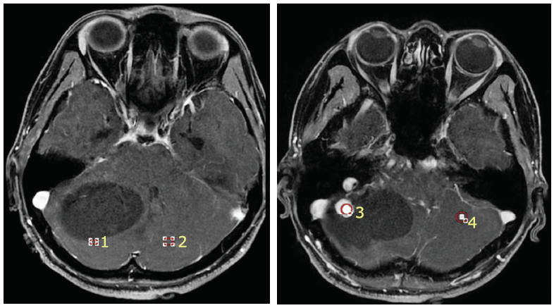
Figure 1.
Figure 2.
MRI of the brain for the patient with hemangioblastoma. The lesion located in the right cerebellum and presented a large cyst with a solid mural nodule. A: axial T2 weighted imaging; B: axial T1 weighted imaging; C: axial enhanced T1 weighted imaging."
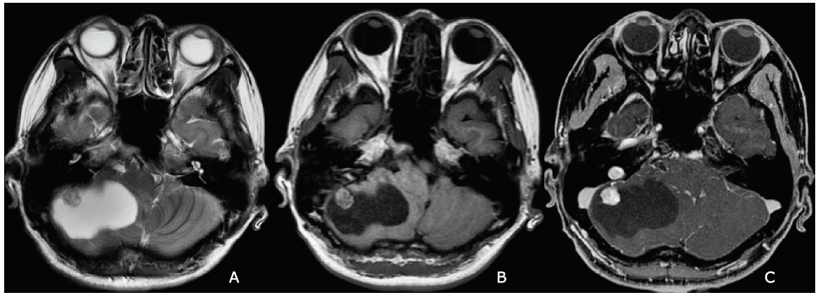
Figure 2.

Figure 3.
Plain and enhanced T2 fluid attenuated inversion recovery (T2-FLAIR) imaging of the brain for the patient with hemangioblastoma."
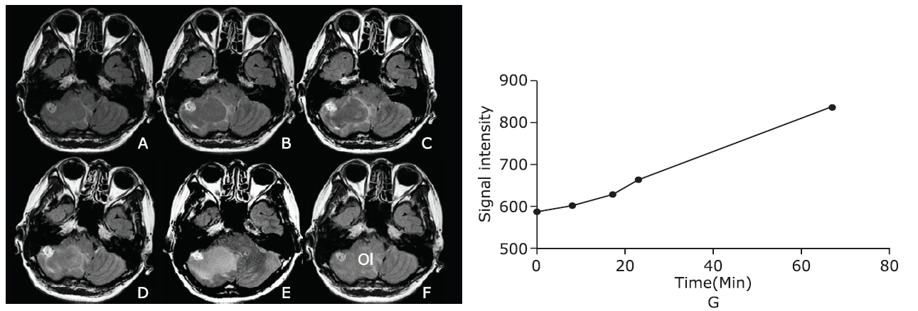
Figure 3.
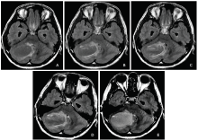
Figure 4.
Plain and enhanced T2-FLAIR imaging of the brain for the patient with hemangioblastoma. The cyst wall of the tumor presented from 5.6 minutes to 23 minutes after contrast administration, and showed no obvious enhancement at 67 minutes after contrast administration. A: plain T2-FLAIR; B: enhanced T2-FLAIR at 5.6 minutes after contrast administration; C: enhanced T2-FLAIR at 17 minutes after contrast administration; D: enhanced T2-FLAIR at 23 minutes after contrast administration; E: enhanced T2-FLAIR at 67 minutes after contrast administration."

Figure 4.
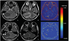
Figure 5.
Ktrans mapping of hemangioblastoma. The enhanced cyst wall on post-contrast T2-FLAIR (B) without enhancement on post-contrast T1 weighted images (A) presented high signal on the Ktrans maps (C). The enhanced mural nodule on post-contrast T1 weighted images (D) without obvious enhancement on T2-FLAIR (E) presented slightly high signal on Ktrans maps (F). Yellow arrows indicate cyst wall on image C and mural nodule on image F."

Figure 5.
"
| Items | Ktrans | Kep | Ve | Vp |
|---|---|---|---|---|
| Cyst wall (ROI-1) | 0.404±0.462 | 0.216±0.270 | 0.634±0.427 | 0.644±0.407 |
| Normal-1 (ROI-2) | 0.028±0.082 | 0.085±0.179 | 0.202±0.270 | 0.793±0.233 |
| Mural nodule (ROI-3) | 0.936±1.854 | 0.708±1.188 | 0.262±0.439 | 0.635±0.483 |
| Normal-2 (ROI-4) | 0.016±0.042 | 0.028±0.096 | 0.218±0.298 | 0.913±0.152 |
| 1. |
Rachinger J, Buslei R, Prell J , et al. Solid haemangioblastomas of the CNS: a review of 17 consecutive cases. Neurosurg Rev 2009; 32(1):37-47. doi: 10.1007/s10143-008-0166-0.
doi: 10.1007/s10143-008-0166-0 pmid: 18810515 |
| 2. |
Wang Z, Hu J, Xu L, Malaguit J , et al. Intratumoral hemorrhage in a patient with cerebellar hemangioblastoma: a case report and review. Medicine 2015; 94(4):e497. doi: 10.1097/MD.0000000000000497.
doi: 10.1097/MD.0000000000000497 |
| 3. |
Chen Z, Liu M, Ma L . Diagnostic value of contrast-enhanced T2-weighted-Fluid-Attenuated Inversion Recovery MR imaging of cerebellar hemangioblastoma: a case report. Zhongguo Yi Xue Ke Xue Yuan Xue Bao 2017; 39(2):296-9. doi: 10.3881/j.issn.1000-503X.2017.02.022.
doi: 10.3881/j.issn.1000-503X.2017.02.022 |
| 4. |
Khalifa F, Soliman A, El-Baz A , et al. Models and methods for analyzing DCE-MRI: a review. Med Phys 2014; 41(12):124301. doi: 10.1118/1.4898202.
doi: 10.1118/1.4898202 pmid: 25471985 |
| 5. |
Tofts PS, Kermode AG . Measurement of the blood-brain barrier permeability and leakage space using dynamic MR imaging. 1. Fundamental concepts. Magn Reson Med 1991; 17(2):357-67. doi: 10.1002/mrm.1910170208.
doi: 10.1002/mrm.1910170208 |
| 6. |
Lee SR, Sanches J, Mark AS , et al. Posterior fossa hemangioblastomas: MR imaging. Radiology 1989; 171(2):463-8. doi: 10.1148/radiology.171.2.2704812.
doi: 10.1148/radiology.171.2.2704812 pmid: 2704812 |
| 7. |
Ho VB, Smirniotopoulos JG, Murphy FM , et al. Radiologic-pathologic correlation: hemangioblastoma. AJNR Am J Neuroradiol 1992; 13(5):1343-52.
doi: 10.1148/radiology.158.3.3511498 pmid: 1414827 |
| 8. |
Lee EK, Lee EJ, Kim S , et al. Importance of contrast-enhanced fluid-attenuated inversion recovery magnetic resonance imaging in various intracranial pathologic conditions. Korean J Radiol 2016; 17(1):127-41. doi: 10.3348/kjr.2016.17.1.127.
doi: 10.3348/kjr.2016.17.1.127 |
| [1] | Huizi Gong, Mengyin Wu, Jun Li, Heyi Zheng. The Great Imitator: Atypical Cutaneous Manifestations of Primary Syphilitic Chancre [J]. Chinese Medical Sciences Journal, 2021, 36(4): 279-283. |
| [2] | Pengfei Qu, Baoliang Bai, Ting Duan, Kai Liu, Jinliang Du, Xin Xiong, Penglin Jia, Zhongchun Sun, Puping Lei. Pneumonia, Multiple Pulmonary Infarction and Abscess Caused by a Bamboo Stick Accidentally Piercing into Chest: a Case Misdiagnosed as Pulmonary Tuberculosis [J]. Chinese Medical Sciences Journal, 2021, 36(3): 252-256. |
| [3] | Lianyan Xu, Ke Yan, Le Lu, Weihong Zhang, Xu Chen, Xiaofei Huo, Jingjing Lu. External and Internal Validation of a Computer Assisted Diagnostic Model for Detecting Multi-Organ Mass Lesions in CT images [J]. Chinese Medical Sciences Journal, 2021, 36(3): 210-217. |
| [4] | Dasheng Li,Dawei Wang,Nana Wang,Haiwang Xu,He Huang,Jianping Dong,Chen Xia. An Insight of the First Community Infected COVID-19 Patient in Beijing by Imported Case: Role of Deep Learning-Assisted CT Diagnosis [J]. Chinese Medical Sciences Journal, 2021, 36(1): 66-71. |
| [5] | Wang Xiaolei, Meng Shanshan, Duan Kehang, Hu Yaowei, Wei Feng. Treatment of Retroperitoneal Cavernous Lymphangioma: A Case Report [J]. Chinese Medical Sciences Journal, 2020, 35(3): 283-285. |
| [6] | Wu Ziquan, Zeng Delu, Yao Jiangling, Bian Yangyang, Gu Yuntao, Meng Zhulong, Fu Jian, Peng Lei. Research Progress on Diagnosis and Treatment of Chronic Osteomyelitis [J]. Chinese Medical Sciences Journal, 2019, 34(3): 211-220. |
| [7] | Wang Yingwei, Zhang Xinghua, Wang Botao, Wang Ye, Liu Mengqi, Wang Haiyi, Ye Huiyi, Chen Zhiye. Value of Texture Analysis of Intravoxel Incoherent Motion Parameters in Differential Diagnosis of Pancreatic Neuroendocrine Tumor and Pancreatic Adenocarcinoma [J]. Chinese Medical Sciences Journal, 2019, 34(1): 1-9. |
| [8] | Wang Botao, Liu Mingxia, Chen Zhiye. Differential Diagnostic Value of Texture Feature Analysis of Magnetic Resonance T2 Weighted Imaging between Glioblastoma and Primary Central Neural System Lymphoma [J]. Chinese Medical Sciences Journal, 2019, 34(1): 10-17. |
| [9] | Wang Botao, Fan Wenping, Xu Huan, Li Lihui, Zhang Xiaohuan, Wang Kun, Liu Mengqi, You Junhao, Chen Zhiye. Value of Magnetic Resonance Imaging Texture Analysis in the Differential Diagnosis of Benign and Malignant Breast Tumors [J]. Chinese Medical Sciences Journal, 2019, 34(1): 33-37. |
| [10] | Bai Mingjian, Feng Jing, Liang Guowei. Urinary Myeloperoxidase to Creatinine Ratio as a New Marker for Diagnosis of Urinary Tract Infection [J]. Chinese Medical Sciences Journal, 2018, 33(3): 152-159. |
| [11] | Li Lihui, Huang Houbin, Chen Zhiye. Early Diagnosis of Recurrent Optic Neuritis Using Contrast-Enhanced T2 Fluid-attenuated Inversion Recovery Imaging: a Case Report [J]. Chinese Medical Sciences Journal, 2018, 33(2): 130-134. |
| [12] | Dong Dexin, Ji Zhigang, Li Hanzhong, Yan Weigang, Zhang Yushi. Preliminary Application of WCX Magnetic Bead-Based Matrix-Assisted Laser Desorption Ionization Time-of-Flight Mass Spectrometry in Analyzing the Urine of Renal Clear Cell Carcinoma [J]. Chinese Medical Sciences Journal, 2017, 32(4): 248-252. |
| [13] | Xu Zeng-xiang, Xie Min, Li Xiao-min, Chen Bing, Lu Lin-ming. Clinicopathological and Genetic Study of an Atypical Renal Hemangioblastoma [J]. Chinese Medical Sciences Journal, 2017, 32(3): 206-210. |
| [14] | Li Tao, Zhao Shaohong, Li Jinfeng, Huang Zili, Luo Chuncai, Yang Li. Value of Multi-detector CT in Detection of Isolated Spontaneous Superior Mesenteric Artery Dissection [J]. Chinese Medical Sciences Journal, 2017, 32(1): 28-33. |
| [15] | Ai-chun Liu, Yan-ying Liu, Yan Li, Li Zhang, Zhan-guo Li. Multiple Myeloma Mimicking Spondyloarthritis: a Case Report [J]. Chinese Medical Sciences Journal, 2014, 29(4): 245-247. |
| Viewed | ||||||
|
Full text |
|
|||||
|
Abstract |
|
|||||
|