Chinese Medical Sciences Journal ›› 2020, Vol. 35 ›› Issue (3): 254-261.doi: 10.24920/003727
新型冠状病毒、非典型肺炎冠状病毒及中东呼吸综合征冠状病毒所致肺炎早期肺部CT表现异同:基于系统性回顾对比分析
陈旭,张刚,郝帅营,白林( ),陆菁菁()
- 北京和睦家医院放射科,北京市朝阳区将台路2号,100015
-
收稿日期:2020-02-29出版日期:2020-09-30发布日期:2020-09-25 -
通讯作者:白林,陆菁菁 E-mail:bai.lin@ufh.com.cn;cjr.lujingjing@vip.163.com
Similarities and Differences of Early Pulmonary CT Features of Pneumonia Caused by SARS-CoV-2, SARS-CoV and MERS-CoV: Comparison Based on a Systemic Review
Chen Xu,Zhang Gang,Hao Shuaiying,Bai Lin(),Lu Jingjing()
- Department of Radiology, Beijing United Family Hospital, Beijing 100015, China
-
Received:2020-02-29Published:2020-09-30Online:2020-09-25 -
Contact:Bai Lin,Lu Jingjing E-mail:bai.lin@ufh.com.cn;cjr.lujingjing@vip.163.com
摘要:
目的 利用系统性回顾分析对比由新型冠状病毒、非典型肺炎冠状病毒及中东呼吸综合征冠状病毒导致的3种病毒性肺炎早期肺部CT表现的相似性及差异性。
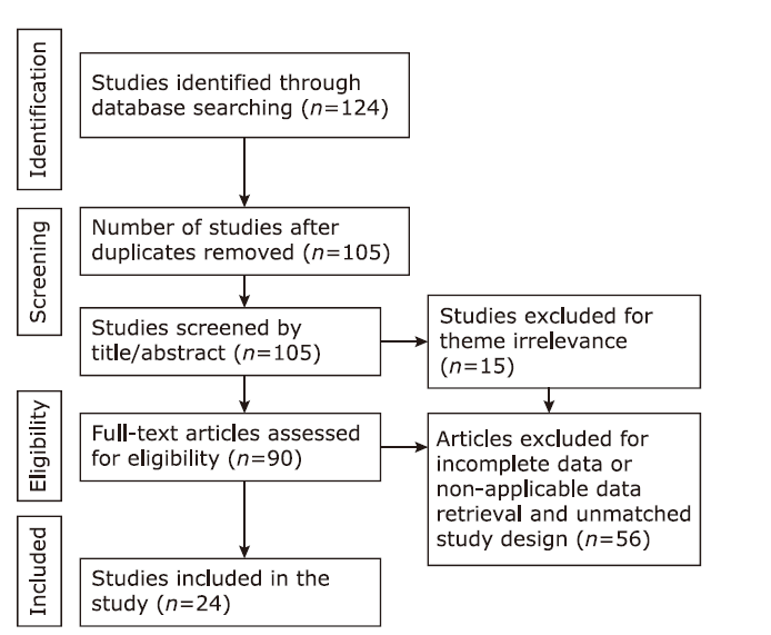
方法 运用电子数据库分别检索成人新型冠状病毒肺炎、非典型肺炎及中东呼吸综合征冠状病毒肺炎CT表现的研究论文及病例报道。文献质量及数据完整性依3位放射科医生所达成的共识进行评估。采用选票计数法纳入各组病例。3组肺炎患者早期胸部CT表现包括病变形态、分布及特殊影像学征象,数据分别提取并记录。利用SPSS 22.0对数据进行对比分析。
结果 本文共纳入24项研究,其中新型冠状病毒病10项、中东呼吸综合征5项以及严重急性呼吸系统综合症9项。纳入的CT检查分别为147、40及122例。对于3种肺炎的早期CT表现,3组间“混合磨玻璃影及实变”、“以磨玻璃影为主”或“以实变为主”的基本病变形态无明显差异(χ2=7.966, P>0.05)。对于“主要累及双肺下叶及胸膜下区”这一病变分布特征,3组间无显著差异(χ 2=4.809, P>0.05)。新型冠状病毒肺炎早期病灶表现更为局限(χ 2=23.509, P<0.05)。铺路石征(χ 2=23.037, P<0.001)、机化性肺炎(P<0.05)及胸腔积液(P<0.001)在新型冠状病毒肺炎的比例明显低于另外两组。尽管纤维条索在全部3种病毒性肺炎中很少出现,但更多见于严重急性呼吸系统综合症(χ 2=6.275, P<0.05)。对于其他影像学表现,只有中东呼吸综合征肺炎早期CT出现了树芽征、空洞,并且其小叶间隔增厚发生率较另外两种肺炎明显增加(χ 2=22.412, P<0.05)。每组早期CT表现均未见气胸、纵隔气肿及淋巴结肿大。
结论 3种冠状病毒肺炎早期影像表现具有相似的基本病变特征,包括磨玻璃影及实变、双侧分布,特别是以双肺下叶及胸膜下区分布为主。新型冠状病毒肺炎早期炎症程度较轻,早期纤维化改变仅见于严重急性呼吸系统综合症。中东呼吸综合征具有更严重的炎性改变包括空洞及胸腔积液。这些差异可能提示每种冠状病毒肺炎具有其特殊的病理生理学进程。
引用本文
Chen Xu, Zhang Gang, Hao Shuaiying, Bai Lin, Lu Jingjing. Similarities and Differences of Early Pulmonary CT Features of Pneumonia Caused by SARS-CoV-2, SARS-CoV and MERS-CoV: Comparison Based on a Systemic Review[J].Chinese Medical Sciences Journal, 2020, 35(3): 254-261.
"
"
| References | Year | Country | Publication type | Sample size (n) |
|---|---|---|---|---|
| COVID-19 | ||||
| Xie et al.[ | 2020 | China | Case reports | 5 |
| Lei et al.[ | 2020 | China | Case report | 11 |
| Fang et al.[ | 2020 | China | Case report | 2 |
| Shi et al.[ | 2020 | China | Case report | 1 |
| Pan et al.[ | 2020 | China | Article | 52 |
| Lin et al.[ | 2020 | China | Case report | 2 |
| Fang et al.[ | 2020 | China | Article | 51 |
| Pan et al.[ | 2020 | China | Article | 21 |
| Duan et al.[ | 2020 | China | Case report | 1 |
| Fang et al.[ | 2020 | China | Case report | 1 |
| MERS | ||||
| Ajlan et al.[ | 2015 | Saudi Arabia | Case reports | 7 |
| Das et al.[ | 2016 | Saudi Arabia | Article | 15 |
| Choi et al.[ | 2016 | Korea | Case report | 1 |
| Hamimi et al.[ | 2015 | Saudi Arabia | Article | 8 |
| Das et al.[ | 2017 | Saudi Arabia | Article | 9 |
| SARS | ||||
| Zhao et al.[ | 2003 | China | Article | 8 |
| Goh et al.[ | 2003 | Singapore | Case report | 1 |
| Nicolaou et al.[ | 2003 | Canada | Case report | 1 |
| Wang et al.[ | 2003 | China | Article | 112 |
| Müller et al.[ | 2003 | Canada | Article | 12 |
| Zeng et al.[ | 2003 | China | Article | 61 |
| Ooi et al.[ | 2003 | Hong Kong | Article | 30 |
| Chan et al.[ | 2004 | Hong Kong | Article | 27 |
| Müller et al.[ | 2004 | Canada | Article | 29 |
"
| Disease category | Number of included studies (n) | Number of included patients (n) | Mean age (yrs) | Early phase CT* (n) |
|---|---|---|---|---|
| COVID-19 | 10 | 147 | 44.8 | 147 |
| MERS | 5 | 40 | 44.8 | 37 |
| SARS | 9 | 122 | 40.4 | 85 |
"
| Items | COVID-19 | MERS | SARS | χ2 | P |
|---|---|---|---|---|---|
| Total number of early phase CT | 147 | 37 | 85 | ||
| GGO consolidation pattern | 7.966 | 0.093 | |||
| Mixed GGO&Consolidation | 41(27.9) | 10(27.0) | 23(27.1) | ||
| GGO mainly | 89(60.5) | 16(43.2) | 49(57.6) | ||
| Consolidation mainly | 17(11.6) | 11(29.7) | 13(15.3) | ||
| Laterality | 13.053 | 0.316 | |||
| Bilaterality | 98(66.7) | 25(67.6) | 39(78.0) | ||
| Unilaterality | 49(33.3) | 12(32.4) | 11(22.0) | ||
| Not accessible* (n) | 0 | 0 | 35 | ||
| More severe in the lower lobes | 4.809 | 0.535 | |||
| Yes | 60(74.7) | 27(73.0) | 11(61.1) | ||
| No | 21(25.9) | 10(27.0) | 7(38.9) | ||
| Not accessible* (n) | 66 | 0 | 67 | ||
| Geographic distribution | 23.509 | <0.05 | |||
| Focal | 45(40.5) | 6(16.2) | 18(42.8) | ||
| Multifocal | 66(59.5) | 20(54.0) | 16(38.1) | ||
| Extensive | 0 | 11(29.7) | 8(19.1) | ||
| Not accessible* (n) | 36 | 0 | 43 | ||
| Transverse distribution | 13.053 | <0.001 | |||
| Central | 15(17.8) | 8(21.6) | 3(4.7) | COVID-19 vs. SARS, P<0.001 | |
| Peripheral | 55(65.4) | 17(45.9) | 28(44.4) | COVID-19 vs. MERS, P=0.09 | |
| No predilection | 14(16.7) | 12(32.4) | 32(50.8) | SARS vs. MERS, P=0.02 | |
| Not accessible* (n) | 63 | 0 | 22 | ||
| Nodular lesion | 0 | 4(10.8) | 14(15.7) | <0.001 | |
| Septal thickening | 17(11.6) | 16(43.2) | 12(13.5) | 22.412 | <0.001 |
| Fibrotic changes | 2(1.4) | 2(5.4) | 7(7.9) | 6.275 | <0.05 |
| Tree-in-bud | 0 | 5(13.5) | 0 | <0.05 | |
| Cavitation | 0 | 4(10.8) | 0 | <0.05 | |
| Crazy paving pattern | 10(6.8) | 10(27.0) | 26(29.2) | 23.037 | <0.001 |
| Organizing pneumonia pattern | 1(0.7) | 4(10.8) | 7(7.9) | <0.05 | |
| Pleural effusion | 0(0) | 21(56.8) | 12(13.5) | <0.001 |
| 1. |
Wu F, Zhao S, Yu B, et al. A new coronavirus associated with human respiratory disease in China. Nature 2020; 579(7798):265-9. doi: 10.1038/s41586-020-2008-3.
doi: 10.1038/s41586-020-2008-3 pmid: 32015508 |
| 2. |
Huang C, Wang Y, Li X, et al. Clinical features of patients infected with 2019 novel coronavirus in Wuhan, China. Lancet 2020; 395(10223):497-506. doi: 10.1016/S0140-6736(20)30183-5.
doi: 10.1016/S0140-6736(20)30183-5 pmid: 31986264 |
| 3. |
Ajlan AM. Reply to “Chest CT findings in MERS”. AJR Am J Roentgenol 2015; 204(1):W112. doi: 10.2214/AJR.14.13367.
doi: 10.2214/AJR.14.13367 pmid: 25539266 |
| 4. |
Xie X, Zhong Z, Zhao W, et al. Chest CT for typical 2019-nCoV pneumonia: relationship to negative RT-PCR testing. Radiology 2020; 296(2):E41-E5. doi: 10.1148/radiol.2020200343.
doi: 10.1148/radiol.2020200343 pmid: 32049601 |
| 5. |
Lei J, Li J, Li X, et al. CT imaging of the 2019 novel coronavirus (2019-nCoV) pneumonia. Radiology 2020; 295(1):18. doi: 10.1148/radiol.2020200236.
doi: 10.1148/radiol.2020200236 pmid: 32003646 |
| 6. |
Fang Y, Zhang H, Xu Y, et al. CT manifestations of two cases of 2019 novel coronavirus (2019-nCoV) pneumonia. Radiology 2020; 295(1):208-9. doi: 10.1148/radiol.2020200280.
pmid: 32031481 |
| 7. | Shi H, Han X, Zheng C. Evolution of CT manifestations in a patient recovered from 2019 novel coronavirus (2019-nCoV) pneumonia in Wuhan, China. Radiology 2020; 295(1):20. doi: 10.1148/radiol.2020200269. |
| 8. | Pan Y, Guan H, Zhou S, et al. Initial CT findings and temporal changes in patients with the novel coronavirus pneumonia (2019-nCoV): a study of 63 patients in Wuhan, China. Eur Radiol 2020; 30(6):3306-9. doi: 10.1007/s00330-020-06731-x. |
| 9. | Lin X, Gong Z, Xiao Z, et al. Novel coronavirus pneumonia outbreak in 2019: computed tomographic findings in two cases. Korean J Radiol 2020; 21(3):365-8. doi: 10.3348/kjr.2020.0078. |
| 10. |
Fang Y, Zhang H, Xie J, et al. Sensitivity of chest CT for COVID-19: comparison to RT-PCR. Radiology 2020; 296(2):E115-E7. doi: 10.1148/radiol.2020200432.
doi: 10.1148/radiol.2020200432 pmid: 32073353 |
| 11. | Pan F, Ye T, Sun P, et al. Time course of lung changes on chest CT during recovery from 2019 novel coronavirus (COVID-19) pneumonia. Radiology 2020; 295(3):715-21. doi: 10.1148/radiol.2020200370. |
| 12. |
Duan YN, Qin J. Pre- and posttreatment chest CT findings: 2019 novel coronavirus (2019-nCoV) pneumonia. Radiology 2020; 295(1):21. doi: 10.1148/radiol.2020200323.
pmid: 32049602 |
| 13. | Fang X, Zhao M, Li S, et al. Changes of CT findings in a 2019 novel coronavirus (2019-nCoV) pneumonia patient. QJM 2020; 113(4):271-2. doi: 10.1093/qjmed/hcaa038. |
| 14. |
Shi H, Han X, Jiang N, et al. Radiological findings from 81 patients with COVID-19 pneumonia in Wuhan, China: a descriptive study. Lancet Infect Dis 2020; 20(4):425-34. doi: 10.1016/S1473-3099(20)30086-4.
doi: 10.1016/S1473-3099(20)30086-4 pmid: 32105637 |
| 15. | Ren LL, Wang YM, Wu ZQ, et al. Identification of a novel coronavirus causing severe pneumonia in human: a descriptive study. Chin Med J (Engl) 2020; 133(9):1015-24. doi: 10.1097/CM9.0000000000000722. |
| 16. | Liu J, Zheng X, Tong Q, et al. Overlapping and discrete aspects of the pathology and pathogenesis of the emerging human pathogenic coronaviruses SARS-CoV, MERS-CoV, and 2019-nCoV. J Med Virol 2020; 92(5):491-4. doi: 10.1002/jmv.25709. |
| 17. |
Xu Z, Shi L, Wang Y, et al. Pathological findings of COVID-19 associated with acute respiratory distress syndrome. Lancet Respir Med 2020; 8(4):420-2. doi: 10.1016/S2213-2600(20)30076-X.
doi: 10.1016/S2213-2600(20)30076-X pmid: 32085846 |
| 18. |
Koo HJ, Lim S, Choe J, et al. Radiographic and CT features of viral pneumonia. Radiographics 2018; 38(3):719-39. doi: 10.1148/rg.2018170048.
pmid: 29757717 |
| 19. |
Chan MS, Chan IY, Fung KH, et al. High-resolution CT findings in patients with severe acute respiratory syndrome: a pattern-based approach. AJR Am J Roentgenol 2004; 182(1):49-56. doi: 10.2214/ajr.182.1.1820049.
pmid: 14684511 |
| 20. | Hansell DM, Bankier AA, MacMahon H, et al. Fleischner Society: glossary of terms for thoracic imaging. Radiology 2008; 246(3):697-722. doi: 10.1148/radiol.2462070712. |
| 21. |
Ooi GC, Khong PL, Muller NL, et al. Severe acute respiratory syndrome: temporal lung changes at thin-section CT in 30 patients. Radiology 2004; 230(3):836-44. doi: 10.1148/radiol.2303030853.
pmid: 14990845 |
| 22. | Das KM, Lee EY, Langer RD, et al. Middle East respiratory syndrome coronavirus: what does a radiologist need to know? AJR Am J Roentgenol 2016; 206(6):1193-201. doi: 10.2214/AJR.15.15363. |
| 23. | Choi WJ, Lee KN, Kang EJ, et al. Middle East respiratory syndrome-coronavirus infection: a case report of serial computed tomographic findings in a young male patient. Korean J Radiol 2016; 17(1):166-70. doi: 10.3348/kjr.2016.17.1.166. |
| 24. | Hamimi A. MERS-CoV: Middle East respiratory syndrome corona virus: can radiology be of help? Initial single center experience. Egyptian J Radiol Nucl Med 2015; 47(1):95-106. doi: 10.1016/j.ejrnm.2015.11.004. |
| 25. |
Das KM, Lee EY, Singh R, et al. Follow-up chest radiographic findings in patients with MERS-CoV after recovery. Indian J Radiol Imaging 2017; 27(3):342-9. doi: 10.4103/ijri.IJRI_469_16.
doi: 10.4103/ijri.IJRI_469_16 pmid: 29089687 |
| 26. | Zhao DW, Ma DQ, Wei W, et al. Early X-ray and CT appearances of severe acute respiratory syndrome an analysis of 28 cases. Chin Med J 2003; 116(6):823-6. |
| 27. |
Goh SK, Tsou YY, Kaw JL. Severe acute respiratory syndrome (SARS): imaging findings during the acute and recovery phases of disease. J Thorac Imaging 2003; 18(3):195-9. doi: 10.1097/00005382-200307000-00010.
pmid: 12867818 |
| 28. |
Nicolaou S, Al-Nakshabandi NA, Müller NL. SARS: imaging of severe acute respiratory syndrome. AJR Am J Roentgenol 2003; 180(5):1247-9. doi: 10.2214/ajr.180.5.1801247.
pmid: 12704032 |
| 29. | Wang R, Sun H, Song L, et al. Plain radiograph and CT features of 112 patients with SARS in acute stage. Beijing Da Xue Xue Bao Yi Xue Ban 2003; 35(Suppl):29-33. |
| 30. |
Müller N, Ooi G, Khong P, et al. Severe acute respiratory syndrome: radiographic and CT findings. AJR Am J Roentgenol 2003; 181(1):3-8.
doi: 10.2214/ajr.181.1.1810003 pmid: 12818821 |
| 31. |
Zeng QS, Chen L, Hu WQ, et al. Roentgenography and CT appearance in patients with severe acute respiratory syndrome. Zhonghua Jie He He Hu Xi Za Zhi 2003; 26(6):347-49.
pmid: 12899767 |
| 32. | Müller NL, Ooi GC, Khong PL, et al. High-resolution CT findings of severe acute respiratory syndrome at presentation and after admission. AJR Am J Roentgenol 2004; 182(1):39-44. doi: 10.2214/ajr.182.1.1820039. |
| 33. | Chen H, Ai L, Lu H, et al. Clinical and imaging features of COVID-19. Radiol Infect Dis 2020; 27(2):43-50. doi: 10.1016/j.jrid.2020.04.003. |
| 34. |
Kim EA, Lee KS, Primack SL, et al. Viral pneumonias in adults: radiologic and pathologic findings. Radiographics 2002; 22 Spec No: S137-49. doi: 10.1148/radiographics.22.suppl_1.g02oc15s137.
pmid: 12376607 |
| 35. |
Wang C, Horby PW, Hayden FG, et al. A novel coronavirus outbreak of global health concern. Lancet 2020; 395(10223):470-3. doi: 10.1016/S0140-6736(20)30185-9.
pmid: 31986257 |
| 36. |
Li Y, Xia L. Coronavirus Disease 2019 (COVID-19): role of chest CT in diagnosis and management. AJR Am J Roentgenol 2020; 214(6):1280-6. doi: 10.2214/AJR.20.22954.
doi: 10.2214/AJR.20.22954 pmid: 32130038 |
| 37. | Wang CH, Liu CY, Wan YL, et al. Persistence of lung inflammation and lung cytokines with high-resolution CT abnormalities during recovery from SARS. Respir Res 2005; 6(1):42. doi: 10.1186/1465-9921-6-42. |
| 38. |
Chang YC, Yu CJ, Chang SC, et al. Pulmonary sequelae in convalescent patients after severe acute respiratory syndrome: evaluation with thin-section CT. Radiology 2005; 236(3):1067-75. doi: 10.1148/radiol.2363040958.
pmid: 16055695 |
| 39. |
Xie L, Liu Y, Xiao Y, et al. Follow-up study on pulmonary function and lung radiographic changes in rehabilitating severe acute respiratory syndrome patients after discharge. Chest 2005; 127(6):2119-24. doi: 10.1378/chest.127.6.2119.
pmid: 15947329 |
| 40. |
Alsaad KO, Hajeer AH, Al BM, et al. Histopathology of Middle East respiratory syndrome coronovirus (MERS-CoV) infection—clinicopathological and ultrastructural study. Histopathology 2018; 72(3):516-24. doi: 10.1111/his.13379.
doi: 10.1111/his.13379 pmid: 28858401 |
| [1] | 杜方智, 张瑞丽, 王千秋. 消除梅毒母婴传播:中国在COVID-19大流行前和流行期间的实践经验[J]. Chinese Medical Sciences Journal, 2022, 37(1): 67-72. |
| [2] | 阿泰菲•贝吉•霍扎尼, 阿米尔穆罕默德•梅拉吉哈, 马赫迪耶•苏莱曼尼. 嗅觉缺失的新冠肺炎患者嗅球的磁共振成像结果:系统综述[J]. Chinese Medical Sciences Journal, 2022, 37(1): 23-30. |
| [3] | 瞿鹏飞, 白宝良, 段婷, 刘凯, 杜进良, 熊鑫, 贾彭林, 孙仲春, 雷普平. 竹签意外刺入胸腔引发的肺炎、多发肺梗塞和肺脓肿:一例误诊为肺结核的病例报告[J]. Chinese Medical Sciences Journal, 2021, 36(3): 252-256. |
| [4] | 宋兰, 朱振宸, 赵瑞杰, 李鹏昌, 田杜雪, 杜铁宽, 徐燕, 杨启文, 曹玮, 宋伟, 金征宇. 北京市单中心19例COVID-19患者的流行病学特征、影像学表现及临床转归[J]. Chinese Medical Sciences Journal, 2021, 36(2): 85-96. |
| [5] | 吴斌,周江华,汪文鑫,杨慧琳,夏盟,张丙宏,折志刚,李红良. 高脂血症与新型冠状病毒肺炎住院患者28天全因死亡率的关系[J]. Chinese Medical Sciences Journal, 2021, 36(1): 17-26. |
| [6] | 李大胜,王大为,王娜娜,徐海旺,黄河,董建平,夏晨. 北京首例由输入病例导致社区感染的新型冠状病毒肺炎患者:深度学习CT辅助诊断的作用[J]. Chinese Medical Sciences Journal, 2021, 36(1): 66-71. |
| [7] | 具杨花, 乔红梅, 张影, 李亚男. 3例病毒相关性儿童横纹肌溶解症的临床分析与文献复习[J]. Chinese Medical Sciences Journal, 2020, 35(4): 383-386. |
| [8] | Vahid Damanpak Moghadam,Hamed Shafiee,Maryam Ghorbani,Reza Heidarifar. 新型冠状肺炎患者气管插管的补充建议[J]. Chinese Medical Sciences Journal, 2020, 35(2): 110-111. |
| [9] | 田毅, 龚亚红, 柳培雨, 王晟, 徐宵寒, 王晓月, 黄宇光. 新型冠状病毒肺炎流行期间围术期感染的防控策略[J]. Chinese Medical Sciences Journal, 2020, 35(2): 114-120. |
| [10] | 刘秉楠, 齐莹. EB病毒感染后瑞氏综合征伴多器官功能障碍的一例重症病例[J]. Chinese Medical Sciences Journal, 2019, 34(4): 297-299. |
| [11] | 吴焱, 宋歌, 魏春波, 伦文辉. 人类免疫缺陷病毒-1感染急性期双侧面神经麻痹合并脑膜炎1例[J]. Chinese Medical Sciences Journal, 2019, 34(1): 55-59. |
| [12] | 欧琴, 李文仿, 李蓓, 于春芳. 耐碳青霉烯类肺炎克雷白杆菌临床分布及整合素1与其耐药关系的研究[J]. Chinese Medical Sciences Journal, 2017, 32(2): 107-112. |
| 阅读次数 | ||||||
|
全文 |
|
|||||
|
摘要 |
|
|||||
|