Chinese Medical Sciences Journal ›› 2021, Vol. 36 ›› Issue (2): 85-96.doi: 10.24920/003821
• Original Article • Previous Articles Next Articles
Epidemiologic Features, Radiological Findings andClinical Outcomes of 19 Patients with COVID-19in a Single Center in Beijing, China
Lan Song1, Zhenchen Zhu1, 2, Ruijie Zhao1, Pengchang Li3, Duxue Tian1, Tiekuan Du4, Yan Xu5, Qiwen Yang3, Wei Cao6, Wei Song1, *( ), Zhengyu Jin1, *()
- 1Department of Radiology,Peking Union Medical College Hospital, Chinese Academy of Medical Sciences & Peking Union Medical College, Beijing 100730, China
2MD Program, Chinese Academy of Medical Sciences & Peking Union Medical College, Beijing 100730, China
3Department of Laboratory Medicine,Peking Union Medical College Hospital, Chinese Academy of Medical Sciences & Peking Union Medical College, Beijing 100730, China
4Department of Emergency,Peking Union Medical College Hospital, Chinese Academy of Medical Sciences & Peking Union Medical College, Beijing 100730, China
5Department of Respiratory and Critical Care Medicine, Peking Union Medical College Hospital, Chinese Academy of Medical Sciences & Peking Union Medical College, Beijing 100730, China
6Department of Infectious Disease, Peking Union Medical College Hospital, Chinese Academy of Medical Sciences & Peking Union Medical College, Beijing 100730, China
-
Received:2021-01-11Published:2021-06-30Online:2021-05-31 -
Contact:Wei Song,Zhengyu Jin E-mail:cjr.songwei@vip.163.com;jinzy@pumch.cn
Cite this article
Lan Song, Zhenchen Zhu, Ruijie Zhao, Pengchang Li, Duxue Tian, Tiekuan Du, Yan Xu, Qiwen Yang, Wei Cao, Wei Song, Zhengyu Jin. Epidemiologic Features, Radiological Findings andClinical Outcomes of 19 Patients with COVID-19in a Single Center in Beijing, China[J].Chinese Medical Sciences Journal, 2021, 36(2): 85-96.
share this article
Add to citation manager EndNote|Reference Manager|ProCite|BibTeX|RefWorks
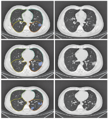
Figure 1.
Examples of pulmonary lobe segmentation and lesion segmentation. Chest CT images of 40-year-old male (case 17) diagnosed with COVID-19. Right: Original axial unenhanced CT images; Left: Pulmonary lobes and lesion segmentation (corresponding output with deep learning algorithm shows the multifocal subpleurally distributed GGOs in left lower lobe). GGO: ground glass opacity."

Figure 1.
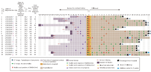
Figure 2.
Time course of symptoms, hospital admission, and discharge of patients infected with SARS-CoV-2. The epidemiological and clinical characteristics of the 19 patients with COVID-19. The cases were successively listed according to the date of diagnosis. Date of diagnosis was defined as the origin point; the contact history was reviewed; abnormal blood test and CT image upon admission were listed; and symptom and nucleic acid test were recorded during hospitalization."
Figure 2.
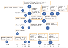
Figure 3.
Epidemic map of 19 COVID-19 patients. Dotted lines indicate close contact between cases. Dates refer to the dates of symptom onset (for 3 asymptomatic patients: case 10 refers to the date arriving in Beijing; case 11 and case 19 refer to the date contacting with case 10)."

Figure 3.
Table 1
Summary of clinical features and laboratory results of patients infected with SARS-CoV-2 at initial presentation (n=19) "
| Items | Results |
|---|---|
| Age [mean±SD (range), yrs] | 42±18 (7-68) |
| Male [No. (%)] | 11 (57.9) |
| Any comorbidity [No. (%)]a | 4 (21.1) |
| Signs and symptoms on presentation [No. (%)] | |
| Fever | 15 (78.9) |
| Dry cough | 11 (57.9) |
| Chill | 6 (31.6) |
| Fatigue | 4 (21.1) |
| Sore throat | 3 (15.8) |
| Myalgia | 3 (15.8) |
| Rhinorrhea | 3 (15.8) |
| Shortness of breath | 2 (10.5) |
| Diarrhea | 2 (10.5) |
| Expectoration | 1 (5.3) |
| Headache | 1 (5.3) |
| Asymptomatic and afebrile | 2 (10.5) |
| Vital signs at presentation [median (range)] | |
| Temperature (℃) | 38 (37.3, 38.0) |
| Pulse oximeter O2 saturation (%) | 98 (97, 99) |
| Baseline investigations [median (range)] | |
| White blood cells [×109/L, mean±SD (range), normal range (3.5-9.5)] | 5.0±1.6 (3.2-10.3) |
| Hemoglobin [g/L, mean±SD (range), normal range (120-160)] | 143.2±21.2 (98-196) |
| Platelets [×109/L, mean±SD (range), normal range (100-350)] | 212±60 (122-336) |
| Neutrophils [×109/L, normal range (2.0-7.5)] | 3.0 (2.0-3.3) |
| Lymphocytes [×109/L, normal range (0.8-4.0)] | 1.5 (0.3-3.3) |
| C-reactive protein [No. (%) above normal range, normal range (0.0-8.0) mg/L] | 11 (57.9) |
| Procalcitonin [No. (%) above normal range, normal range <0.5 ng/ml, n=15] | 1 (6.7) |
| LDH [No. (%) above normal range, normal range (0-250) U/L, n=9] | 1 (11.1) |
| Creatine kinase [No.(%) above normal range, normal range (24-195) U/L] | 3 (15.8) |
| Cardiac troponin I [No. (%) above normal range, normal range (0-0.04) μg/L, n=13] | 1 (7.7) |
| NT-proBNP [No. (%) of negative results, normal range (0-125) g/ml, n=12] | 12 (100) |
| CK-MB [No. (%) of negative results, normal range (0-3.6) μg/L, n=13] | 13 (100) |
| Abnormal chest CT [No. (%)] | 16 (84.2) |
| Duration of symptoms [d, median (P25, P75), n=17] | 12 (10, 13.5) |
| Days of hospitalization [d, mean±SD (range)] | 20±5 (11-25) |
Table 1
Table 2
Baseline CT imaging findings in 262 lesions of 19 patients with COVID-19"
| CT imaging findings | No. of patients (%) | No. of lesions (%) |
|---|---|---|
| n | 19 | 262 |
| Distribution | ||
| Peripheral distribution | 14 (73.7) | 223 (85.1) |
| Both peripheral and central distribution | 2 (10.5) | 39 (14.9) |
| Peri-bronchial distribution | 14 (73.7) | 170 (64.9) |
| Anterior predominant | 1 (5.3) | 65 (24.8) |
| Posterior predominant | 15 (78.9) | 197 (75.2) |
| Upper lobe distribution | 14 (73.7) | 80 (30.5) |
| Middle lobe distribution | 9 (47.4) | 31 (11.8) |
| Lower lobe distribution | 14 (73.7) | 151 (57.6) |
| Patterns of the lesion | ||
| Pure ground glass opacities | 15 (78.9) | 87 (33.2) |
| Ground glass opacities with consolidation | 15 (78.9) | 147 (56.1) |
| Consolidation | 7 (36.8) | 28 (10.7) |
| Other signs surrounding the lesion | ||
| Halo-sign | 8 (42.1) | 69 (26.3) |
| Reversed halo-sign | 3 (15.8) | 18 (6.9) |
| Other signs in the lesion | ||
| Interlobular septal thickening | 10 (52.6) | 102 (38.9) |
| Crazy paving pattern | 7 (36.8) | 30 (11.5) |
| Air bronchogram sign | 15 (78.9) | 89 (34.0) |
| Architectural deformation | 6 (31.6) | 36 (13.7) |
| Bronchus distortion | 6 (31.6) | 38 (14.5) |
| Bronchial wall thickening | 13 (68.4) | 76 (29.0) |
| Vascular dilation sign | 16 (84.2) | 105 (40.1) |
| Vacuole sign | 7 (36.8) | 38 (14.5) |
Table 2
Table 3
Lobe of lesion distribution of the pulmonary involvement in each lobe in 19 patients of COVID-19"
| Lobe of lesion distribution | No. (%) of 19 patients | Percentage of total 57 involved lobes (%) |
|---|---|---|
| Left upper lobe | 11 (57.9) | 11 (19.3) |
| Left lower lobe | 14 (73.7) | 14 (24.6) |
| Right upper lobe | 11 (57.9) | 11 (19.3) |
| Right middle lobe | 9 (47.4) | 9 (15.8) |
| Right lower lobe | 12 (63.2) | 12 (21.1) |
Table 3
Table 4
Baseline quantitative CT parameters in the initial CT-positive 16 patients with COVID-19 and 5 family clusters (including 13 patients) [mean±SD (range)]"
| Clusters | n | No. of lesions (n) | Total lesion (volume, ml) | Mean CT value (HU) | Pneumonia volume/total lung volume (%) |
|---|---|---|---|---|---|
| CT positive patients | 16 | 13.8±17.8 (1-53) | 216.7±292.5 (2.7-944.7) | -357.2±166.9 (-583.3--135.4) | 6.4±10.6 (0.1-40) |
| Family cluster 1 | 2 | 9.0±1.4 (8-10) | 320.9±335.5 (83.7-558.1) | -534.6±43.8 (-565.6--506.6) | 7.1±5.9 (2.9-11.2) |
| Family cluster 2 | 2 | 19.5±13.4 (10-29) | 116.6±145.7 (13.6-219.6) | -394.6±166.9 (-470.7--318.4) | 4.3±5.8 (0.2-8.4) |
| Family cluster 3 | 4 | 6.5±4.7 (0-10) | 452.7±457.5 (0-944.7) | -275.0±173.8 (-475.3--164.5) | 15.5±18.0 (0-40) |
| Family cluster 4 | 2 | 2.0±2.8 (0-4) | 3.8±5.4 (0-7.6) | -466.4±68.7 (-515.0--417.8) | 0.1±0.1 (0-0.2) |
| Family cluster 5 | 3 | 8.0±8.5 (0-17) | 13.4±16.7 (0-32.1) | -238.2±127.6 (-328.4--148.0) | 0.3±0.3 (0-0.6) |
Table 4
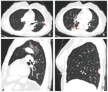
Figure 4.
Thin-slice CT images in three patients with COVID-19. A. 60-year-old woman (case 4). CT image obtained on day 3 after onset of symptoms shows subpleural patchy GGO in the lingual segment of the left upper lobe, with vascular dilation (arrow). B. The same patient on the same day. CT shows subpleural patchy consolidation with scattered vacuole signs (arrow) and bronchial wall thickening (arrowhead) in the dorsal segment of the right lower lobe. C. 68-year-old man (case 5, husband of case 4). CT image obtained on day 19 after onset of symptoms shows multiple patchy GGOs with predominant peri-bronchial distribution and multiple vacuole signs (arrow) in the left upper lobe. D. 31-year-old woman (case 6). CT image obtained on day 4 after onset of symptoms shows subpleural wedge-shaped consolidation with vascular dilation (arrowhead) and fuzzy margin in the right lower lobe."
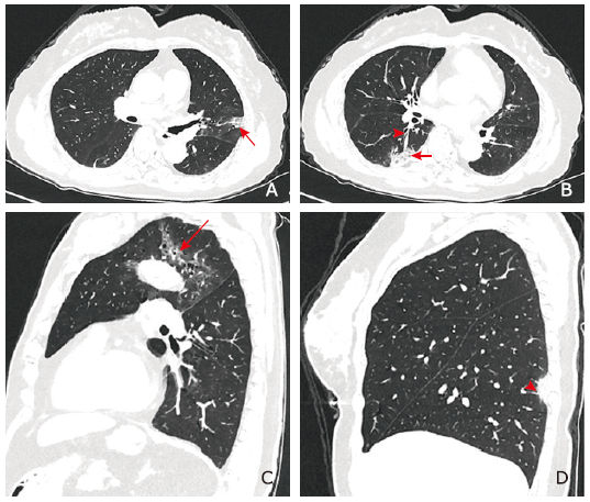
Figure 4.

Figure 5.
Thin-slice CT images in three patients of a family with COVID-19. A. 61-year-old woman (case 10). CT image obtained on day 15 after the date on which she arrived in Beijing shows diffuse subpleural and peri-bronchial confluent GGOs and fibrous cords in both lungs with architecture deformation (arrow) and crazy paving sign (box). B. 64-year-old man (case 18, husband of case 10). CT image obtained on day 15 after the date on which he arrived in Beijing shows diffuse subpleural confluent GGOs and mixed GGOs with consolidations accompanied by crazy paving sign and traction bronchiectasis in both lungs (box). C. Asymptomatic and afebrile 37-year-old woman (case 19, daughter of case 10 and case 18). Her nucleic acid test results of SARS-CoV-2 were negative for 6 times. The seventh nucleic acid test result was positive after 30 days from her first test. CT image obtained on day 14 after contacting with case 10 shows a subpleural patchy consolidations with halo sign (box) in the right lower lobe."
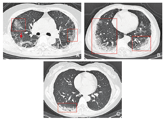
Figure 5.
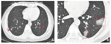
Figure 6.
Thin-slice CT images in two patients with COVID-19. A. Afebrile 39-year-old woman (case 15, wife of case 17). CT image obtained on day 9 after onset of symptoms shows scattered pure GGO nodule (arrow) or part-solid nodule (arrowhead) distributed along the bronchovascular bundle in the bilateral lower lobes. B. 40-year-old man (case 17). CT image obtained on day 5 after onset of symptoms shows subpleural scattered GGOs with crazy paving sign in left lower lobe (box)."

Figure 6.
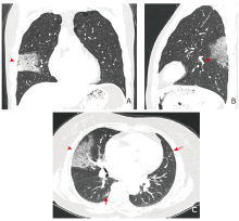
Figure 7.
Thin-slice CT images in three patients with COVID-19. A. 35-year-old man (case 2). CT image obtained on day 10 after onset of symptoms shows large segmental distribution GGO (arrowhead) in the lateral segment of right middle lobe. B. 42-year-old man (case 1). CT image obtained on day 4 after onset of symptoms shows large segmental distribution GGO (arrowhead) in the dorsal segment of left lower lobe. C. 63-year-old woman (case 9). CT image obtained on day 3 after onset of symptoms shows large segmental distribution GGO (arrowhead) in the right middle lobe and multiple subpleural little patchy GGOs (arrow) in the right lower lobe and the left lung."

Figure 7.
Figure 8.
Serial CT images in a 62-year-old male with COVID-19. A. The initial CT image obtained on day 3 after onset of symptoms shows round GGO in left lower lobe. B. After 3 days, the first repeat CT shows irregular GGO with heterogeneous density and increasing in extent and density. C, D. Subsequently, lesions absorb gradually with decrease of extent and density (C, 11 days later), and only leaves a small amount of faint GGO on the latest CT scan (D, 16 days later)."
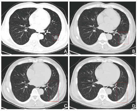
Figure 8.
| [1.] |
Huang C, Wang Y, Li X, et al. Clinical features of patients infected with 2019 novel coronavirus in Wuhan, China. Lancet 2020; 395(10223):497-506. doi: 10.1016/S0140-6736(20)30183-5.
doi: 10.1016/S0140-6736(20)30183-5 |
| [2.] |
Wang D, Hu B, Hu C, et al. Clinical characteristics of 138 hospitalized patients with 2019 novel coronavirus-infected pneumonia in Wuhan, China. JAMA 2020; 323(11):1061-9. doi: 10.1001/jama.2020.1585.
doi: 10.1001/jama.2020.1585 |
| [3.] |
Chan JF, Yuan S, Kok KH, et al. A familial cluster of pneumonia associated with the 2019 novel coronavirus indicating person-to-person transmission: a study of a family cluster. Lancet 2020; 395(10223):514-23. doi: 10.1016/S0140-6736(20)30154-9.
doi: 10.1016/S0140-6736(20)30154-9 |
| [4.] |
Pan Y, Guan H, Zhou S, et al. Initial CT findings and temporal changes in patients with the novel coronavirus pneumonia (2019-nCoV): a study of 63 patients in Wuhan, China. Eur Radiol 2020; 30(6):3306-9. doi: 10.1007/s00330-020-06731-x.
doi: 10.1007/s00330-020-06731-x |
| [5.] |
Zhu N, Zhang D, Wang W, et al. A novel coronavirus from patients with pneumonia in China, 2019. N Engl J Med 2020; 382(8):727-33. doi: 10.1056/NEJMoa2001017.
doi: 10.1056/NEJMoa2001017 |
| [6.] | World Health Organization. Coronavirus disease 2019 (COVID-19) Situation Report. 2021; https://www.who.int/publications/m/item/weekly-epidemiological-update—5-january-2021. Accessed January 5, 2021. |
| [7.] |
Corman VM, Landt O, Kaiser M, et al. Detection of 2019 novel coronavirus (2019-nCoV) by real-time RT-PCR. Euro Surveill 2020; 25(3):2000045. doi: 10.2807/1560-7917.ES.2020.25.3.2000045.
doi: 10.2807/1560-7917.ES.2020.25.3.2000045 |
| [8.] |
Wang C, Horby PW, Hayden FG, et al. A novel coronavirus outbreak of global health concern. Lancet 2020; 395(10223):470-3. doi: 10.1016/S0140-6736(20)30185-9.
doi: 10.1016/S0140-6736(20)30185-9 |
| [9.] | National Health Commision of China. COVID-19 epidemiological distribution. 2020; http://2019ncov.chinacdc.cn/2019-nCoV/. Accessed January 5, 2021. |
| [10.] |
Shi H, Han X, Jiang N, et al. Radiological findings from 81 patients with COVID-19 pneumonia in Wuhan, China: a descriptive study. Lancet Infect Dis 2020; 20(4):425-34. doi: 10.1016/S1473-3099(20)30086-4.
doi: 10.1016/S1473-3099(20)30086-4 |
| [11.] |
Tian S, Hu N, Lou J, et al. Characteristics of COVID-19 infection in Beijing. J Infect 2020; 80(4):401-6. doi: 10.1016/j.jinf.2020.02.018.
doi: 10.1016/j.jinf.2020.02.018 |
| [12.] |
Hu Z, Song C, Xu C, et al. Clinical characteristics of 24 asymptomatic infections with COVID-19 screened among close contacts in Nanjing, China. Sci China Life Sci 2020; 63(5):706-11. doi: 10.1007/s11427-020-1661-4.
doi: 10.1007/s11427-020-1661-4 |
| [13.] |
Lai CC, Liu YH, Wang CY, et al. Asymptomatic carrier state, acute respiratory disease, and pneumonia due to severe acute respiratory syndrome coronavirus 2 (SARS-CoV-2): facts and myths. J Microbiol Immunol Infect 2020; 53(3):404-12. doi: 10.1016/j.jmii.2020.02.012.
doi: 10.1016/j.jmii.2020.02.012 |
| [14.] |
Chen N, Zhou M, Dong X, et al. Epidemiological and clinical characteristics of 99 cases of 2019 novel coronavirus pneumonia in Wuhan, China: a descriptive study. Lancet 2020; 395(10223):507-13. doi: 10.1016/S0140-6736(20)30211-7.
doi: 10.1016/S0140-6736(20)30211-7 |
| [15.] |
Xia W, Shao J, Guo Y, et al. Clinical and CT features in pediatric patients with COVID-19 infection: different points from adults. Pediatr Pulmonol 2020; 55(5):1169-74. doi: 10.1002/ppul.24718.
doi: 10.1002/ppul.24718 |
| [16.] |
Young BE, Ong SWX, Kalimuddin S, et al. Epidemiologic features and clinical course of patients infected with SARS-CoV-2 in Singapore. JAMA 2020; 323(15):1488-94. doi: 10.1001/jama.2020.3204.
doi: 10.1001/jama.2020.3204 |
| [17.] | China National Health Commission. Diagnosis and treatment of pneumonia caused by novel coronavirus (trial version 7). 2020; http://www.nhc.gov.cn/yzygj/s7653p/202003/46c9294a7dfe4cef80dc7f5912eb1989/files/ce3e6945832a438eaae415350a8ce964.pdf |
| [18.] | Li ZH, Zhang S, Zhang JG, et al. MVP-Net: multi-view FPN with position-aware attention for deep universal lesion detection. In: Shen D, et al. (eds) Medical Image Computing and Computer Assisted Intervention-MICCAI 2019. MICCAI 2019. Lecture Notes in Computer Science, vol 11769. Springer, Cham. https://doi.org/10.1007/978-3-030-32226-7_2 |
| [19.] |
Chang D, Lin M, Wei L, et al. Epidemiologic and clinical characteristics of novel coronavirus infections involving 13 patients outside Wuhan, China. JAMA 2020; 323(11):1092-3. doi: 10.1001/jama.2020.1623.
doi: 10.1001/jama.2020.1623 pmid: 32031568 |
| [20.] |
Chung M, Bernheim A, Mei X, et al. CT imaging features of 2019 novel coronavirus (2019-nCoV). Radiology 2020; 295(1):202-7. doi: 10.1148/radiol.2020200230.
doi: 10.1148/radiol.2020200230 |
| [21.] |
Pan F, Ye T, Sun P, et al. Time course of lung changes on chest CT during recovery from 2019 novel coronavirus (COVID-19) pneumonia. Radiology 2020; 295(3):200370. doi: 10.1148/radiol.2020200370.
doi: 10.1148/radiol.2020200370 |
| [22.] |
Wong KT, Antonio GE, Hui DS, et al. Thin-section CT of severe acute respiratory syndrome: evaluation of 73 patients exposed to or with the disease. Radiology 2003; 228(2):395-400. doi: 10.1148/radiol.2283030541.
doi: 10.1148/radiol.2283030541 pmid: 12738877 |
| [23.] |
Das KM, Lee EY, Enani MA, et al. CT correlation with outcomes in 15 patients with acute Middle East respiratory syndrome coronavirus. AJR Am J Roentgenol 2015; 204(4):736-42. doi: 10.2214/AJR.14.13671.
doi: 10.2214/AJR.14.13671 |
| [24.] |
Li Y, Xia L. Coronavirus disease 2019 (COVID-19): role of chest CT in diagnosis and management. AJR Am J Roentgenol 2020; 214(6):1280-6. doi: 10.2214/AJR.20.22954.
doi: 10.2214/AJR.20.22954 |
| [25.] |
Ai T, Yang Z, Hou H, et al. Correlation of chest CT and RT-PCR testing in coronavirus disease 2019 (COVID-19) in China: a report of 1014 cases. Radiology 2020; 296(2):E32-E40. doi: 10.1148/radiol.2020200642.
doi: 10.1148/radiol.2020200642 |
| [26.] |
Zou L, Ruan F, Huang M, et al. SARS-CoV-2 viral load in upper respiratory specimens of infected patients. N Engl J Med 2020; 382(12):1177-9. doi: 10.1056/NEJMc2001737.
doi: 10.1056/NEJMc2001737 |
| [27.] |
Zhou Y, Yang GD, Feng K, et al. Clinical features and chest CT findings of coronavirus disease 2019 in infants and young children. Zhongguo Dang Dai Er Ke Za Zhi 2020; 22(3):215-20. doi: 10.7499/j.issn.1008-8830.2020.03.007.
doi: 10.7499/j.issn.1008-8830.2020.03.007 |
| [28.] | Lytvynenko J. The first infant has died in the US after testing positive for the coronavirus. 2020; https://www.buzzfeednews.com/article/janelytvynenko/baby-us-dies-covid19-coronavirus. Accessed March 30, 2020. |
| [29.] | Dursun A. Iran: 6-year-old child dies from coronavirus. 2020; https://www.aa.com.tr/en/latest-on-coronavirus-outbreak/iran-6-year-old-child-dies-from-coronavirus/1777045#. Accessed March 30, 2020. |
| [30.] |
Lan L, Xu D, Ye G, et al. Positive RT-PCR test results in patients recovered from COVID-19. JAMA 2020; 323(15):1502-3. doi: 10.1001/jama.2020.2783.
doi: 10.1001/jama.2020.2783 pmid: 32105304 |
| [31.] |
Rothe C, Schunk M, Sothmann P, et al. Transmission of 2019-nCoV infection from an asymptomatic contact in Germany. N Engl J Med 2020; 382(10):970-1. doi: 10.1056/NEJMc2001468.
doi: 10.1056/NEJMc2001468 |
| [32.] |
Wu JT, Leung K, Leung GM. Nowcasting and forecasting the potential domestic and international spread of the 2019-nCoV outbreak originating in Wuhan, China: a modelling study. Lancet 2020; 395(10225):689-97. doi: 10.1016/S0140-6736(20)30260-9.
doi: 10.1016/S0140-6736(20)30260-9 |
| [1] | Changyi Liu, Xiaoqing Liu, Xiaochun Shi. Clinical Features of Spontaneous Remission in the Classic Fever of Unknown Origin: A Retrospective Study [J]. Chinese Medical Sciences Journal, 2022, 37(2): 134-141. |
| [2] | Fangzhi Du, Ruili Zhang, Qianqiu Wang. Eliminating Mother-to-Child Transmission of Syphilis: Chinese Practice before and during COVID-19 Pandemics [J]. Chinese Medical Sciences Journal, 2022, 37(1): 67-72. |
| [3] | Atefeh Beigi-khoozani, Amirmohammad Merajikhah, Mahdieh Soleimani. Magnetic Resonance Imaging Findings of Olfactory Bulb in Anosmic Patients with COVID-19: A Systematic Review [J]. Chinese Medical Sciences Journal, 2022, 37(1): 23-30. |
| [4] | Bin Wu,Jianghua Zhou,Wenxin Wang,Huilin Yang,Meng Xia,Binghong Zhang,Zhigang She,Hongliang Li. Association Analysis of Hyperlipidemia with the 28-Day All-Cause Mortality of COVID-19 in Hospitalized Patients [J]. Chinese Medical Sciences Journal, 2021, 36(1): 17-26. |
| [5] | Dasheng Li,Dawei Wang,Nana Wang,Haiwang Xu,He Huang,Jianping Dong,Chen Xia. An Insight of the First Community Infected COVID-19 Patient in Beijing by Imported Case: Role of Deep Learning-Assisted CT Diagnosis [J]. Chinese Medical Sciences Journal, 2021, 36(1): 66-71. |
| [6] | Jian Cao, Guorong Wang, Zhiwei Wang, Zhengyu Jin. CT Texture Analysis: A Potential Biomarker for Evaluating KRAS Mutational Status in Colorectal Cancer [J]. Chinese Medical Sciences Journal, 2020, 35(4): 306-314. |
| [7] | Chen Xu, Zhang Gang, Hao Shuaiying, Bai Lin, Lu Jingjing. Similarities and Differences of Early Pulmonary CT Features of Pneumonia Caused by SARS-CoV-2, SARS-CoV and MERS-CoV: Comparison Based on a Systemic Review [J]. Chinese Medical Sciences Journal, 2020, 35(3): 254-261. |
| [8] | Zuo Mingzhang,Huang Yuguang,Ma Wuhua,Xue Zhanggang,Zhang Jiaqiang,Gong Yahong,Che Lu, Chinese Society of Anesthesiology Task Force on Airway Management. Expert Recommendations for Tracheal Intubation in Critically Ill Patients with Noval Coronavirus Disease 2019 [J]. Chinese Medical Sciences Journal, 2020, 35(2): 105-109. |
| [9] | Damanpak Moghadam Vahid,Shafiee Hamed,Ghorbani Maryam,Heidarifar Reza. Letter to the Editor: Additional Recommendations before Intubation of COVID-19 Patients [J]. Chinese Medical Sciences Journal, 2020, 35(2): 110-111. |
| [10] | Tian Yi, Gong Yahong, Liu Peiyu, Wang Sheng, Xu Xiaohan, Wang Xiaoyue, Huang Yuguang. Infection Prevention Strategy in Operating Room during Coronavirus Disease 2019 (COVID-19) Outbreak [J]. Chinese Medical Sciences Journal, 2020, 35(2): 114-120. |
| [11] | Li Ping, Zhu Liang, Wang Xuan, Xue Huadan, Wu Xin, Jin Zhengyu. Imaging Diagnosis of Type Ⅲ Choledochal Cyst: A Case Report [J]. Chinese Medical Sciences Journal, 2018, 33(3): 194-203. |
| [12] | Li Tao, Yang Li, Zhang Weiguo, Luo Chuncai, Huang Zili, Li Jinfeng, Li Xin. Midterm Follow-up of Coronary Artery Bypass Grafting with 64-Slice Multi-detector Computed Tomography: Identification of Risk Factors Affecting Graft Patency [J]. Chinese Medical Sciences Journal, 2018, 33(2): 69-76. |
| [13] | Wang Ting, Ma Lin, Lou Xin, Bu Bo. Trigeminal Ganglioneuroma in the Middle-posterior Cranial Fossa: a Case Report△ [J]. Chinese Medical Sciences Journal, 2017, 32(2): 123-128. |
| [14] | Li Tao, Zhao Shaohong, Li Jinfeng, Huang Zili, Luo Chuncai, Yang Li. Value of Multi-detector CT in Detection of Isolated Spontaneous Superior Mesenteric Artery Dissection [J]. Chinese Medical Sciences Journal, 2017, 32(1): 28-33. |
| [15] | Chen Xing, Wang Lihua, Zhang Lixian, Zhao Caihong. IgG4-related Autoimmune Pancreatitis Mimicking Acute Pancreatitis: A Case Report [J]. Chinese Medical Sciences Journal, 2017, 32(1): 65-68. |
| Viewed | ||||||
|
Full text |
|
|||||
|
Abstract |
|
|||||
|