Chinese Medical Sciences Journal ›› 2020, Vol. 35 ›› Issue (3): 254-261.doi: 10.24920/003727
• Original Article • Previous Articles Next Articles
Similarities and Differences of Early Pulmonary CT Features of Pneumonia Caused by SARS-CoV-2, SARS-CoV and MERS-CoV: Comparison Based on a Systemic Review
Chen Xu, Zhang Gang, Hao Shuaiying, Bai Lin( ), Lu Jingjing()
- Department of Radiology, Beijing United Family Hospital, Beijing 100015, China
-
Received:2020-02-29Published:2020-09-30Online:2020-09-25 -
Contact:Bai Lin,Lu Jingjing E-mail:bai.lin@ufh.com.cn;cjr.lujingjing@vip.163.com
Cite this article
Chen Xu, Zhang Gang, Hao Shuaiying, Bai Lin, Lu Jingjing. Similarities and Differences of Early Pulmonary CT Features of Pneumonia Caused by SARS-CoV-2, SARS-CoV and MERS-CoV: Comparison Based on a Systemic Review[J].Chinese Medical Sciences Journal, 2020, 35(3): 254-261.
share this article
Add to citation manager EndNote|Reference Manager|ProCite|BibTeX|RefWorks
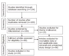
Figure 1.
Flow diagram of study selection."
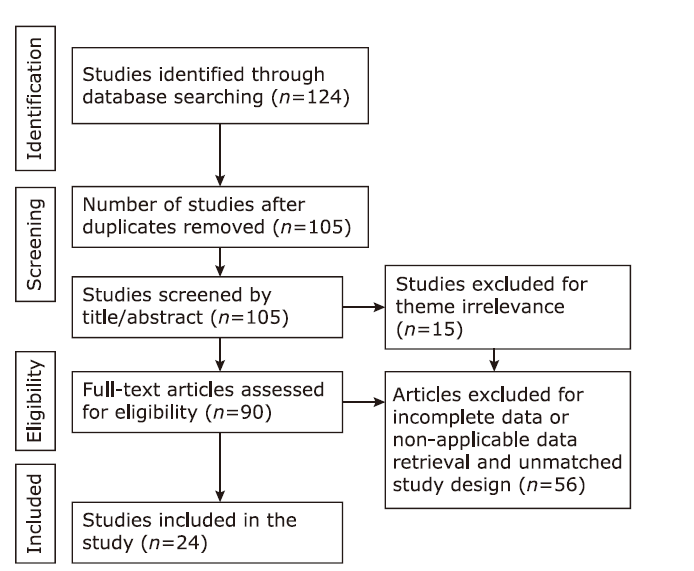
Figure 1.
Table 1
Main characteristics of the included studies"
| References | Year | Country | Publication type | Sample size (n) |
|---|---|---|---|---|
| COVID-19 | ||||
| Xie et al.[ | 2020 | China | Case reports | 5 |
| Lei et al.[ | 2020 | China | Case report | 11 |
| Fang et al.[ | 2020 | China | Case report | 2 |
| Shi et al.[ | 2020 | China | Case report | 1 |
| Pan et al.[ | 2020 | China | Article | 52 |
| Lin et al.[ | 2020 | China | Case report | 2 |
| Fang et al.[ | 2020 | China | Article | 51 |
| Pan et al.[ | 2020 | China | Article | 21 |
| Duan et al.[ | 2020 | China | Case report | 1 |
| Fang et al.[ | 2020 | China | Case report | 1 |
| MERS | ||||
| Ajlan et al.[ | 2015 | Saudi Arabia | Case reports | 7 |
| Das et al.[ | 2016 | Saudi Arabia | Article | 15 |
| Choi et al.[ | 2016 | Korea | Case report | 1 |
| Hamimi et al.[ | 2015 | Saudi Arabia | Article | 8 |
| Das et al.[ | 2017 | Saudi Arabia | Article | 9 |
| SARS | ||||
| Zhao et al.[ | 2003 | China | Article | 8 |
| Goh et al.[ | 2003 | Singapore | Case report | 1 |
| Nicolaou et al.[ | 2003 | Canada | Case report | 1 |
| Wang et al.[ | 2003 | China | Article | 112 |
| Müller et al.[ | 2003 | Canada | Article | 12 |
| Zeng et al.[ | 2003 | China | Article | 61 |
| Ooi et al.[ | 2003 | Hong Kong | Article | 30 |
| Chan et al.[ | 2004 | Hong Kong | Article | 27 |
| Müller et al.[ | 2004 | Canada | Article | 29 |
Table 1
Table 2
Basic results of the pooled study and CT availability information"
| Disease category | Number of included studies (n) | Number of included patients (n) | Mean age (yrs) | Early phase CT* (n) |
|---|---|---|---|---|
| COVID-19 | 10 | 147 | 44.8 | 147 |
| MERS | 5 | 40 | 44.8 | 37 |
| SARS | 9 | 122 | 40.4 | 85 |
Table 2
Table 3
Comparison of imaging features and patterns of early phase CT for COVID-19, MERS and SARS [n (%)]"
| Items | COVID-19 | MERS | SARS | χ2 | P |
|---|---|---|---|---|---|
| Total number of early phase CT | 147 | 37 | 85 | ||
| GGO consolidation pattern | 7.966 | 0.093 | |||
| Mixed GGO&Consolidation | 41(27.9) | 10(27.0) | 23(27.1) | ||
| GGO mainly | 89(60.5) | 16(43.2) | 49(57.6) | ||
| Consolidation mainly | 17(11.6) | 11(29.7) | 13(15.3) | ||
| Laterality | 13.053 | 0.316 | |||
| Bilaterality | 98(66.7) | 25(67.6) | 39(78.0) | ||
| Unilaterality | 49(33.3) | 12(32.4) | 11(22.0) | ||
| Not accessible* (n) | 0 | 0 | 35 | ||
| More severe in the lower lobes | 4.809 | 0.535 | |||
| Yes | 60(74.7) | 27(73.0) | 11(61.1) | ||
| No | 21(25.9) | 10(27.0) | 7(38.9) | ||
| Not accessible* (n) | 66 | 0 | 67 | ||
| Geographic distribution | 23.509 | <0.05 | |||
| Focal | 45(40.5) | 6(16.2) | 18(42.8) | ||
| Multifocal | 66(59.5) | 20(54.0) | 16(38.1) | ||
| Extensive | 0 | 11(29.7) | 8(19.1) | ||
| Not accessible* (n) | 36 | 0 | 43 | ||
| Transverse distribution | 13.053 | <0.001 | |||
| Central | 15(17.8) | 8(21.6) | 3(4.7) | COVID-19 vs. SARS, P<0.001 | |
| Peripheral | 55(65.4) | 17(45.9) | 28(44.4) | COVID-19 vs. MERS, P=0.09 | |
| No predilection | 14(16.7) | 12(32.4) | 32(50.8) | SARS vs. MERS, P=0.02 | |
| Not accessible* (n) | 63 | 0 | 22 | ||
| Nodular lesion | 0 | 4(10.8) | 14(15.7) | <0.001 | |
| Septal thickening | 17(11.6) | 16(43.2) | 12(13.5) | 22.412 | <0.001 |
| Fibrotic changes | 2(1.4) | 2(5.4) | 7(7.9) | 6.275 | <0.05 |
| Tree-in-bud | 0 | 5(13.5) | 0 | <0.05 | |
| Cavitation | 0 | 4(10.8) | 0 | <0.05 | |
| Crazy paving pattern | 10(6.8) | 10(27.0) | 26(29.2) | 23.037 | <0.001 |
| Organizing pneumonia pattern | 1(0.7) | 4(10.8) | 7(7.9) | <0.05 | |
| Pleural effusion | 0(0) | 21(56.8) | 12(13.5) | <0.001 |
Table 3
| 1. |
Wu F, Zhao S, Yu B, et al. A new coronavirus associated with human respiratory disease in China. Nature 2020; 579(7798):265-9. doi: 10.1038/s41586-020-2008-3.
doi: 10.1038/s41586-020-2008-3 pmid: 32015508 |
| 2. |
Huang C, Wang Y, Li X, et al. Clinical features of patients infected with 2019 novel coronavirus in Wuhan, China. Lancet 2020; 395(10223):497-506. doi: 10.1016/S0140-6736(20)30183-5.
doi: 10.1016/S0140-6736(20)30183-5 pmid: 31986264 |
| 3. |
Ajlan AM. Reply to “Chest CT findings in MERS”. AJR Am J Roentgenol 2015; 204(1):W112. doi: 10.2214/AJR.14.13367.
doi: 10.2214/AJR.14.13367 pmid: 25539266 |
| 4. |
Xie X, Zhong Z, Zhao W, et al. Chest CT for typical 2019-nCoV pneumonia: relationship to negative RT-PCR testing. Radiology 2020; 296(2):E41-E5. doi: 10.1148/radiol.2020200343.
doi: 10.1148/radiol.2020200343 pmid: 32049601 |
| 5. |
Lei J, Li J, Li X, et al. CT imaging of the 2019 novel coronavirus (2019-nCoV) pneumonia. Radiology 2020; 295(1):18. doi: 10.1148/radiol.2020200236.
doi: 10.1148/radiol.2020200236 pmid: 32003646 |
| 6. |
Fang Y, Zhang H, Xu Y, et al. CT manifestations of two cases of 2019 novel coronavirus (2019-nCoV) pneumonia. Radiology 2020; 295(1):208-9. doi: 10.1148/radiol.2020200280.
pmid: 32031481 |
| 7. | Shi H, Han X, Zheng C. Evolution of CT manifestations in a patient recovered from 2019 novel coronavirus (2019-nCoV) pneumonia in Wuhan, China. Radiology 2020; 295(1):20. doi: 10.1148/radiol.2020200269. |
| 8. | Pan Y, Guan H, Zhou S, et al. Initial CT findings and temporal changes in patients with the novel coronavirus pneumonia (2019-nCoV): a study of 63 patients in Wuhan, China. Eur Radiol 2020; 30(6):3306-9. doi: 10.1007/s00330-020-06731-x. |
| 9. | Lin X, Gong Z, Xiao Z, et al. Novel coronavirus pneumonia outbreak in 2019: computed tomographic findings in two cases. Korean J Radiol 2020; 21(3):365-8. doi: 10.3348/kjr.2020.0078. |
| 10. |
Fang Y, Zhang H, Xie J, et al. Sensitivity of chest CT for COVID-19: comparison to RT-PCR. Radiology 2020; 296(2):E115-E7. doi: 10.1148/radiol.2020200432.
doi: 10.1148/radiol.2020200432 pmid: 32073353 |
| 11. | Pan F, Ye T, Sun P, et al. Time course of lung changes on chest CT during recovery from 2019 novel coronavirus (COVID-19) pneumonia. Radiology 2020; 295(3):715-21. doi: 10.1148/radiol.2020200370. |
| 12. |
Duan YN, Qin J. Pre- and posttreatment chest CT findings: 2019 novel coronavirus (2019-nCoV) pneumonia. Radiology 2020; 295(1):21. doi: 10.1148/radiol.2020200323.
pmid: 32049602 |
| 13. | Fang X, Zhao M, Li S, et al. Changes of CT findings in a 2019 novel coronavirus (2019-nCoV) pneumonia patient. QJM 2020; 113(4):271-2. doi: 10.1093/qjmed/hcaa038. |
| 14. |
Shi H, Han X, Jiang N, et al. Radiological findings from 81 patients with COVID-19 pneumonia in Wuhan, China: a descriptive study. Lancet Infect Dis 2020; 20(4):425-34. doi: 10.1016/S1473-3099(20)30086-4.
doi: 10.1016/S1473-3099(20)30086-4 pmid: 32105637 |
| 15. | Ren LL, Wang YM, Wu ZQ, et al. Identification of a novel coronavirus causing severe pneumonia in human: a descriptive study. Chin Med J (Engl) 2020; 133(9):1015-24. doi: 10.1097/CM9.0000000000000722. |
| 16. | Liu J, Zheng X, Tong Q, et al. Overlapping and discrete aspects of the pathology and pathogenesis of the emerging human pathogenic coronaviruses SARS-CoV, MERS-CoV, and 2019-nCoV. J Med Virol 2020; 92(5):491-4. doi: 10.1002/jmv.25709. |
| 17. |
Xu Z, Shi L, Wang Y, et al. Pathological findings of COVID-19 associated with acute respiratory distress syndrome. Lancet Respir Med 2020; 8(4):420-2. doi: 10.1016/S2213-2600(20)30076-X.
doi: 10.1016/S2213-2600(20)30076-X pmid: 32085846 |
| 18. |
Koo HJ, Lim S, Choe J, et al. Radiographic and CT features of viral pneumonia. Radiographics 2018; 38(3):719-39. doi: 10.1148/rg.2018170048.
pmid: 29757717 |
| 19. |
Chan MS, Chan IY, Fung KH, et al. High-resolution CT findings in patients with severe acute respiratory syndrome: a pattern-based approach. AJR Am J Roentgenol 2004; 182(1):49-56. doi: 10.2214/ajr.182.1.1820049.
pmid: 14684511 |
| 20. | Hansell DM, Bankier AA, MacMahon H, et al. Fleischner Society: glossary of terms for thoracic imaging. Radiology 2008; 246(3):697-722. doi: 10.1148/radiol.2462070712. |
| 21. |
Ooi GC, Khong PL, Muller NL, et al. Severe acute respiratory syndrome: temporal lung changes at thin-section CT in 30 patients. Radiology 2004; 230(3):836-44. doi: 10.1148/radiol.2303030853.
pmid: 14990845 |
| 22. | Das KM, Lee EY, Langer RD, et al. Middle East respiratory syndrome coronavirus: what does a radiologist need to know? AJR Am J Roentgenol 2016; 206(6):1193-201. doi: 10.2214/AJR.15.15363. |
| 23. | Choi WJ, Lee KN, Kang EJ, et al. Middle East respiratory syndrome-coronavirus infection: a case report of serial computed tomographic findings in a young male patient. Korean J Radiol 2016; 17(1):166-70. doi: 10.3348/kjr.2016.17.1.166. |
| 24. | Hamimi A. MERS-CoV: Middle East respiratory syndrome corona virus: can radiology be of help? Initial single center experience. Egyptian J Radiol Nucl Med 2015; 47(1):95-106. doi: 10.1016/j.ejrnm.2015.11.004. |
| 25. |
Das KM, Lee EY, Singh R, et al. Follow-up chest radiographic findings in patients with MERS-CoV after recovery. Indian J Radiol Imaging 2017; 27(3):342-9. doi: 10.4103/ijri.IJRI_469_16.
doi: 10.4103/ijri.IJRI_469_16 pmid: 29089687 |
| 26. | Zhao DW, Ma DQ, Wei W, et al. Early X-ray and CT appearances of severe acute respiratory syndrome an analysis of 28 cases. Chin Med J 2003; 116(6):823-6. |
| 27. |
Goh SK, Tsou YY, Kaw JL. Severe acute respiratory syndrome (SARS): imaging findings during the acute and recovery phases of disease. J Thorac Imaging 2003; 18(3):195-9. doi: 10.1097/00005382-200307000-00010.
pmid: 12867818 |
| 28. |
Nicolaou S, Al-Nakshabandi NA, Müller NL. SARS: imaging of severe acute respiratory syndrome. AJR Am J Roentgenol 2003; 180(5):1247-9. doi: 10.2214/ajr.180.5.1801247.
pmid: 12704032 |
| 29. | Wang R, Sun H, Song L, et al. Plain radiograph and CT features of 112 patients with SARS in acute stage. Beijing Da Xue Xue Bao Yi Xue Ban 2003; 35(Suppl):29-33. |
| 30. |
Müller N, Ooi G, Khong P, et al. Severe acute respiratory syndrome: radiographic and CT findings. AJR Am J Roentgenol 2003; 181(1):3-8.
doi: 10.2214/ajr.181.1.1810003 pmid: 12818821 |
| 31. |
Zeng QS, Chen L, Hu WQ, et al. Roentgenography and CT appearance in patients with severe acute respiratory syndrome. Zhonghua Jie He He Hu Xi Za Zhi 2003; 26(6):347-49.
pmid: 12899767 |
| 32. | Müller NL, Ooi GC, Khong PL, et al. High-resolution CT findings of severe acute respiratory syndrome at presentation and after admission. AJR Am J Roentgenol 2004; 182(1):39-44. doi: 10.2214/ajr.182.1.1820039. |
| 33. | Chen H, Ai L, Lu H, et al. Clinical and imaging features of COVID-19. Radiol Infect Dis 2020; 27(2):43-50. doi: 10.1016/j.jrid.2020.04.003. |
| 34. |
Kim EA, Lee KS, Primack SL, et al. Viral pneumonias in adults: radiologic and pathologic findings. Radiographics 2002; 22 Spec No: S137-49. doi: 10.1148/radiographics.22.suppl_1.g02oc15s137.
pmid: 12376607 |
| 35. |
Wang C, Horby PW, Hayden FG, et al. A novel coronavirus outbreak of global health concern. Lancet 2020; 395(10223):470-3. doi: 10.1016/S0140-6736(20)30185-9.
pmid: 31986257 |
| 36. |
Li Y, Xia L. Coronavirus Disease 2019 (COVID-19): role of chest CT in diagnosis and management. AJR Am J Roentgenol 2020; 214(6):1280-6. doi: 10.2214/AJR.20.22954.
doi: 10.2214/AJR.20.22954 pmid: 32130038 |
| 37. | Wang CH, Liu CY, Wan YL, et al. Persistence of lung inflammation and lung cytokines with high-resolution CT abnormalities during recovery from SARS. Respir Res 2005; 6(1):42. doi: 10.1186/1465-9921-6-42. |
| 38. |
Chang YC, Yu CJ, Chang SC, et al. Pulmonary sequelae in convalescent patients after severe acute respiratory syndrome: evaluation with thin-section CT. Radiology 2005; 236(3):1067-75. doi: 10.1148/radiol.2363040958.
pmid: 16055695 |
| 39. |
Xie L, Liu Y, Xiao Y, et al. Follow-up study on pulmonary function and lung radiographic changes in rehabilitating severe acute respiratory syndrome patients after discharge. Chest 2005; 127(6):2119-24. doi: 10.1378/chest.127.6.2119.
pmid: 15947329 |
| 40. |
Alsaad KO, Hajeer AH, Al BM, et al. Histopathology of Middle East respiratory syndrome coronovirus (MERS-CoV) infection—clinicopathological and ultrastructural study. Histopathology 2018; 72(3):516-24. doi: 10.1111/his.13379.
doi: 10.1111/his.13379 pmid: 28858401 |
| [1] | Fangzhi Du, Ruili Zhang, Qianqiu Wang. Eliminating Mother-to-Child Transmission of Syphilis: Chinese Practice before and during COVID-19 Pandemics [J]. Chinese Medical Sciences Journal, 2022, 37(1): 67-72. |
| [2] | Atefeh Beigi-khoozani, Amirmohammad Merajikhah, Mahdieh Soleimani. Magnetic Resonance Imaging Findings of Olfactory Bulb in Anosmic Patients with COVID-19: A Systematic Review [J]. Chinese Medical Sciences Journal, 2022, 37(1): 23-30. |
| [3] | Pengfei Qu, Baoliang Bai, Ting Duan, Kai Liu, Jinliang Du, Xin Xiong, Penglin Jia, Zhongchun Sun, Puping Lei. Pneumonia, Multiple Pulmonary Infarction and Abscess Caused by a Bamboo Stick Accidentally Piercing into Chest: a Case Misdiagnosed as Pulmonary Tuberculosis [J]. Chinese Medical Sciences Journal, 2021, 36(3): 252-256. |
| [4] | Lan Song, Zhenchen Zhu, Ruijie Zhao, Pengchang Li, Duxue Tian, Tiekuan Du, Yan Xu, Qiwen Yang, Wei Cao, Wei Song, Zhengyu Jin. Epidemiologic Features, Radiological Findings andClinical Outcomes of 19 Patients with COVID-19in a Single Center in Beijing, China [J]. Chinese Medical Sciences Journal, 2021, 36(2): 85-96. |
| [5] | Bin Wu,Jianghua Zhou,Wenxin Wang,Huilin Yang,Meng Xia,Binghong Zhang,Zhigang She,Hongliang Li. Association Analysis of Hyperlipidemia with the 28-Day All-Cause Mortality of COVID-19 in Hospitalized Patients [J]. Chinese Medical Sciences Journal, 2021, 36(1): 17-26. |
| [6] | Zuo Mingzhang,Huang Yuguang,Ma Wuhua,Xue Zhanggang,Zhang Jiaqiang,Gong Yahong,Che Lu, Chinese Society of Anesthesiology Task Force on Airway Management. Expert Recommendations for Tracheal Intubation in Critically Ill Patients with Noval Coronavirus Disease 2019 [J]. Chinese Medical Sciences Journal, 2020, 35(2): 105-109. |
| [7] | Damanpak Moghadam Vahid,Shafiee Hamed,Ghorbani Maryam,Heidarifar Reza. Letter to the Editor: Additional Recommendations before Intubation of COVID-19 Patients [J]. Chinese Medical Sciences Journal, 2020, 35(2): 110-111. |
| [8] | Tian Yi, Gong Yahong, Liu Peiyu, Wang Sheng, Xu Xiaohan, Wang Xiaoyue, Huang Yuguang. Infection Prevention Strategy in Operating Room during Coronavirus Disease 2019 (COVID-19) Outbreak [J]. Chinese Medical Sciences Journal, 2020, 35(2): 114-120. |
| [9] | Ou Qin, Li Wenfang, Li Bei, Yu Chunfang. Prevalence of Carbapenem-Resistant Klebsiella Pneumoniae (CRKP) and the Distribution of Class 1 Integron in Their Strains Isolated from a Hospital in Central China [J]. Chinese Medical Sciences Journal, 2017, 32(2): 107-112. |
| [10] | Wei Chen, Bei Li, Shuai Li, Yi-wen Ou, Qin Ou. Effects of Scutellaria Baicalensis on Activity and Biofilm Formation of Klebsiella Pneumoniae [J]. Chinese Medical Sciences Journal, 2016, 31(3): 180-184. |
| [11] | Dong-qian Yi, Xue-feng Yang, Duan-fang Liao, Qing Wu, Nian Fu, Yang Hu, Ting Cao. Effect of Autophagy Over Liver Diseases [J]. Chinese Medical Sciences Journal, 2016, 31(1): 65-68. |
| [12] | Hong-min Zhang,Da-wei Liu*,Xiao-ting Wang,Yun Long,Huan Chen. Bloodstream Infection with Carbapenem-resistant Klebsiella Pneumoniae and Multidrug-resistant Acinetobacter Baumannii: a Case Report [J]. Chinese Medical Sciences Journal, 2014, 29(1): 51-54. |
| [13] | Tao Wang, Hua-shan Zhao, Qiu-ling Zhang, Chang-lin Xu, and Chang-bai Liu. Generation of Transgene-free Induced Pluripotent Stem Cells with Non-viral Methods [J]. Chinese Medical Sciences Journal, 2013, 28(1): 50-54. |
| [14] | Yao Zhang, Hua Zhang, Jun Xu, Chan Wu, and Xiao-jun Ma. Lack of Response in Severe Pneumocystis Pneumonia to Combined Caspofungin and Clindamycin Treatment: a Case Report [J]. Chinese Medical Sciences Journal, 2011, 26(4): 246-248. |
| Viewed | ||||||
|
Full text |
|
|||||
|
Abstract |
|
|||||
|