Chinese Medical Sciences Journal ›› 2019, Vol. 34 ›› Issue (2): 110-119.doi: 10.24920/003576
多图谱方法在脑MR图像分割中的应用
孙亮1,张丽2,张道强1,*( )
)
- 1. 南京航空航天大学 计算机科学与技术学院 模式分析与机器智能工业和信息化部重点实验室,南京 211106
2. 南京医科大学附属脑科医院 老年医学科,南京 210029
-
收稿日期:2019-02-27接受日期:2019-04-28出版日期:2019-05-14发布日期:2019-05-16 -
通讯作者:张道强 E-mail:dqzhang@nuaa.edu.cn
Multi-Atlas Based Methods in Brain MR Image Segmentation
Sun Liang1,Zhang Li2,Zhang Daoqiang1,*()
- 1. College of Computer Science and Technology, Nanjing University of Aeronautics and Astronautics, MIIT Key Laboratory of Pattern Analysis and Machine Intelligence, Nanjing 211106, China
2. Department of Geriatrics, the Affiliated Brain Hospital of Nanjing Medical University, Nanjing 210029, China
-
Received:2019-02-27Accepted:2019-04-28Published:2019-05-14Online:2019-05-16 -
Contact:Zhang Daoqiang E-mail:dqzhang@nuaa.edu.cn
摘要:
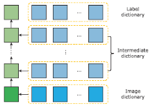
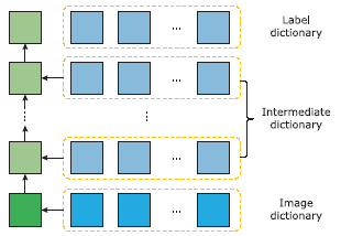
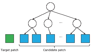
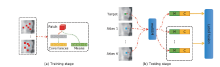
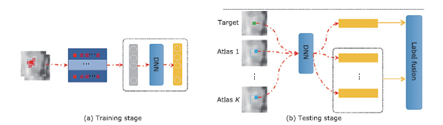
脑感兴趣区域分割是很多脑疾病计算机辅助分析的重要步骤。然而人类大脑具有复杂的解剖结构,同时脑磁共振(Magnetic Resonance, MR)图像处理通常会面临感兴趣区域灰度对比度低,个体之间和个体内的差异性等问题。为了解决这些问题,近年来,基于多图谱的很多方法被用于脑感兴趣区域的分割。在这篇综述中,我们对一些基于多图谱的脑MR图像分割方法进行系统的介绍,包括常用的配准工具箱,经典的标签融合方法,常用数据集以及多图谱分割在临床研究中的应用。我们认为,将图谱图像的解剖结构先验信息融入端到端的深度学习框架中用于脑图像感兴趣区域分割将是多图谱方法未来一个重要的研究方向。
引用本文
Sun Liang,Zhang Li,Zhang Daoqiang. Multi-Atlas Based Methods in Brain MR Image Segmentation[J].Chinese Medical Sciences Journal, 2019, 34(2): 110-119.
"
"
"

"
"
"
"
| Methods | Subjects (n) | DR (%) | ASD | References No |
|---|---|---|---|---|
| LWJoint | 139 | 89.7(88.8) | - | 25 |
| DDLS | 202 | 87.2 | - | 28 |
| HLF | 66 | 88.5 | 0.334 | 32 |
| Progressive SPBL | 64 | 88.3 | - | 35 |
| DSPBL | 66 | 88.3 | 0.40 | 36 |
| NL1 | 135 | 86.45 | - | 38 |
| [1] |
Devanand DP, Pradhaban G, Liu X , et al. Hippocampal and entorhinal atrophy in mild cognitive impairment - Prediction of Alzheimer disease. Neurology 2007; 68(11):828-36. doi:
doi: 10.1212/01.wnl.0000256697.20968.d7 |
| [2] |
Zhang DQ, Wang YP, Zhou LP , et al. Multimodal classification of Alzheimer’s disease and mild cognitive impairment. Neuroimage 2011; 55(3):856-67. doi:
doi: 10.1016/j.neuroimage.2011.01.008 |
| [3] |
Liu MX, Zhang DQ, Shen DG . Relationship induced multi-template learning for diagnosis of Alzheimer’s disease and mild cognitive impairment. IEEE Trans Med Imaging 2016; 35(6):1463-74. doi:
doi: 10.1109/TMI.2016.2515021 |
| [4] |
Jie B, Liu MX, Zhang DQ , et al. Sub-network kernels for measuring similarity of brain connectivity networks in disease diagnosis. IEEE Trans Image Process 2018; 27(5):2340-53. doi:
doi: 10.1109/TIP.2018.2799706 |
| [5] |
Liu MX, Zhang J, Adeli E , et al. Landmark-based deep multi-instance learning for brain disease diagnosis. Med Image Anal 2018; 43:157-68. doi:
doi: 10.1016/j.media.2017.10.005 |
| [6] |
Jenkinson M, Smith S . A global optimisation method for robust affine registration of brain images. Med Image Anal 2001; 5(2):143-56. doi:
doi: 10.1016/S1361-8415(01)00036-6 |
| [7] |
Langerak TR, van der Heide UA, Kotte ANTJ , et al. Improving label fusion in multi-atlas based segmentation by locally combining atlas selection and performance estimation. Comput Vision Image Understand 2015; 130:71-9. doi:
doi: 10.1016/j.cviu.2014.09.004 |
| [8] |
Smith SM, Jenkinson M, Woolrich MW , et al. Advances in functional and structural MR image analysis and implementation as FSL. Neuroimage 2004; 23:S208-S19. doi:
doi: 10.1016/j.neuroimage.2004.07.051 |
| [9] |
Avants BB, Epstein CL, Grossman M , et al. Symmetric diffeomorphic image registration with cross-correlation: Evaluating automated labeling of elderly and neurodegenerative brain. Med Image Anal 2008; 12(1):26-41. doi:
doi: 10.1016/j.media.2007.06.004 |
| [10] |
Vercauteren T, Pennec X, Perchant A , et al. Diffeomorphic demons: Efficient non-parametric image registration. Neuroimage 2009; 45(1):S61-S72. doi:
doi: 10.1016/j.neuroimage.2008.10.040 |
| [11] |
Thirion JP . Image matching as a diffusion process: an analogy with Maxwell’s demons. Med Image Anal 1998; 2(3):243-60. doi:
doi: 10.1016/S1361-8415(98)80022-4 |
| [12] |
Warfield SK, Zou KH, Wells WM . Simultaneous truth and performance level estimation (STAPLE): An algorithm for the validation of image segmentation. IEEE Trans Med Imaging 2004; 23(7):903-21. doi:
doi: 10.1109/TMI.2004.828354 |
| [13] |
Heckemann RA, Hajnal JV, Aljabar P , et al. Automatic anatomical brain MRI segmentation combining label propagation and decision fusion. Neuroimage 2006; 33(1):115-26. doi:
doi: 10.1016/j.neuroimage.2006.05.061 |
| [14] |
Artaechevarria X, Munoz-Barrutia A , Ortiz-de-Solorzano C. Combination strategies in multi-atlas image segmentation: application to brain MR data. IEEE Trans Med Imaging 2009; 28(8):1266-77. doi:
doi: 10.1109/TMI.2009.2014372 |
| [15] |
Aljabar P, Heckemann RA, Hammers A , et al. Multi-atlas based segmentation of brain images: Atlas selection and its effect on accuracy. Neuroimage 2009; 46(3): 726-38. doi: .
doi: 10.1016/j.neuroimage.2009.02.018 |
| [16] |
Lotjonen JMP, Wolz R, Koikkalainen JR , et al. Fast and robust multi-atlas segmentation of brain magnetic resonance images. Neuroimage 2010; 49(3):2352-65. doi:
doi: 10.1016/j.neuroimage.2009.10.026 |
| [17] |
Sabuncu MR, Yeo BTT, Van Leemput K , et al. A Generative model for image segmentation based on label fusion. IEEE Trans Med Imaging 2010; 29(10):1714-29. doi:
doi: 10.1109/TMI.2010.2050897 |
| [18] |
Langerak TR, van der Heide UA, Kotte ANTJ , et al. Label fusion in atlas-based segmentation using a selective and iterative method for performance level estimation (SIMPLE). IEEE Trans Med Imaging 2010; 29(12):2000-8. doi:
doi: 10.1109/TMI.2010.2057442 |
| [19] |
L?tj?nen JM, Wolz R, Koikkalainen JR , et al. Fast and robust multi-atlas segmentation of brain magnetic resonance images. Neuroimage 2010; 49(3):2352-65.
doi: 10.1016/j.neuroimage.2009.10.026 |
| [20] |
Langerak TR, van der Heide VA, Kotte ANTJ , et al. Label fusion in atlas-based segmentation using a selective and iterative method for performance level estimation (SIMPLE). IEEE Trans Med Imaging 2010; 29(12):2000-8. doi:
doi: 10.1109/TMI.2010.2057442 |
| [21] |
Zhang DQ, Wu GR, Jia HJ , et al. Confidence-guided sequential label Fusion for multi-atlas based segmentation. Int Conference on Medical Image Computing and Computer-Assisted Intervention; 2011 September 18-22; Toronto, Canada. Berlin, Heidelberg: Springer; 2011. p 643-50. doi: .
doi: 10.1007/978-3-642-23626-6_79 |
| [22] |
Coupé P, Manjon JV, Fonov V , et al. Patch-based segmentation using expert priors: Application to hippocampus and ventricle segmentation. Neuroimage 2011; 54(2):940-54. doi:
doi: 10.1016/j.neuroimage.2010.09.018 |
| [23] |
Rousseau F, Habas PA, Studholme C . A supervised patch-based approach for human brain labeling. IEEE Trans Med Imaging 2011; 30(10):1852-62. doi:
doi: 10.1109/TMI.2011.2156806 |
| [24] |
Zhang D, Guo Q, Wu G , et al., editors. Sparse patch-based label fusion for multi-atlas segmentation. International Workshop on Multimodal Brain Image Analysis. 2012 Oct 1-5; Nice, France. Berlin, Heidelberg: Springer; 2012. p 94-102. doi: .
doi: 10.1007/978-3-642-33530-3_8 |
| [25] |
Wang HZ, Suh JW, Das SR , et al. Multi-atlas segmentation with joint label fusion. IEEE Trans Pattern Anal Machine Intell 2013; 35(3):611-23. doi:
doi: 10.1109/TPAMI.2012.143 |
| [26] |
Wang HZ, Yushkevich PA . Groupwise segmentation with multi-atlas joint label fusion. International Conference on Medical Image Computing and Computer-Assisted Intervention. 2013 Sep 22-26; Nagoya, Japan. Berlin, Heidelberg: Springer; 2013. p 711-8. doi: .
doi: 10.1007/978-3-642-40811-3_89 |
| [27] |
Wang HZ, Yushkevich PA . Multi-atlas segmentation with joint label fusion and corrective learning — an open source implementation. Front Neuroinform 2013; 7. doi: .
doi: 10.3389/fninf.2013.00027 |
| [28] |
Tong T, Wolz R, Coupe P , et al. Segmentation of MR images via discriminative dictionary learning and sparse coding: Application to hippocampus labeling. Neuroimage 2013; 76(1):11-23. doi:
doi: 10.1016/j.neuroimage.2013.02.069 |
| [29] |
Cardoso MJ, Leung K, Modat M , et al. STEPS: Similarity and Truth Estimation for Propagated Segmentations and its application to hippocampal segmentation and brain parcelation. Med Image Anal 2013; 17(6):671-84. doi:
doi: 10.1016/j.media.2013.02.006 |
| [30] |
Wu GR, Wang Q, Zhang DQ , et al. A generative probability model of joint label fusion for multi-atlas based brain segmentation. Med Image Anal 2014; 18(6):881-90. doi:
doi: 10.1016/j.media.2013.10.013 |
| [31] |
Sun L, Zu C, Zhang D . Reliability guided forward and backward patch-based method for multi-atlas segmentation. International Workshop on Patch-based Techniques in Medical Imaging; 2015 Oct 9; Munich, Germany. Cham: Springer; 2015. p 128-36. doi: .
doi: 10.1007/978-3-319-28194-0_16 |
| [32] |
Wu GR, Kim MJ, Sanroma G , et al. Hierarchical multi-atlas label fusion with multi-scale feature representation and label-specific patch partition. Neuroimage 2015; 106:34-46. doi:
doi: 10.1016/j.neuroimage.2014.11.025 |
| [33] |
Song Y, Wu G, Sun Q , et al. Progressive Label Fusion Framework for Multi-atlas Segmentation by Dictionary Evolution. International Conference on Medical Image Computing and Computer-Assisted Intervention; 2015 October 5-9; Munich, Germany. Cham: Springer; 2015: p 190-7. doi: .
doi: 10.1007/978-3-319-24574-4_23 |
| [34] |
Benkarim OM, Piella G, Ballester MAG , et al. Discriminative confidence estimation for probabilistic multi-atlas label fusion. Med Image Anal 2017; 42:274-87. doi:
doi: 10.1016/j.media.2017.08.008 |
| [35] |
Song YT, Wu GR, Bahrami K , et al. Progressive multi-atlas label fusion by dictionary evolution. Med Image Anal 2017; 36:162-71. doi:
doi: 10.1016/j.media.2016.11.005 |
| [36] |
Zu C, Wang ZX, Zhang DQ , et al. Robust multi-atlas label propagation by deep sparse representation. Pattern Recognition 2017; 63:511-7. doi:
doi: 10.1016/j.patcog.2016.09.028 |
| [37] |
Liang S, Wei S , Zhang D, editors. High-order boltzmann machine-based unsupervised feature learning for multi-atlas segmentation. IEEE International Symposium on Biomedical Imaging. 2017 Apr 18-21; Melbourne, Australia. New York: IEEE; 2017. p 507-10. doi: .
doi: 10.1109/ISBI.2017.7950571 |
| [38] |
Sanroma G, Benkarim OM, Piella G , et al. Learning non-linear patch embeddings with neural networks for label fusion. Med Image Anal 2018; 44:143-55. doi:
doi: 10.1016/j.media.2017.11.013 |
| [39] |
Huo J, Wu J, Cao JW , et al. Supervoxel based method for multi-atlas segmentation of brain MR images. Neuroimage 2018; 175:201-14. doi:
doi: 10.1016/j.neuroimage.2018.04.001 |
| [40] |
Sun L, Zu C, Shao W , et al. Reliability-based robust multi-atlas label fusion for brain MRI segmentation. Artif Intell Med 2019; 96:12-24. doi:
doi: 10.1016/j.artmed.2019.03.004 |
| [41] |
Kittler J . Combining classifiers: A theoretical framework. Pattern Anal App 1998; 1(1):18-27. doi:
doi: 10.1007/BF01238023 |
| [42] |
Wang H, Yushkevich P . Multi-atlas segmentation with joint label fusion and corrective learning—an open source implementation. Front Neuroinform 2013; 7:27. doi: .
doi: 10.3389/fninf.2013.00027 |
| [43] |
Zhang QA, Li BX . Discriminative K-SVD for Dictionary Learning in Face Recognition. IEEE Conference on Computer Vision and Pattern Recognition. 2010 Jun 13-18; San Francisco, CA, USA. New York: IEEE; 2010. p 2691-8. doi: .
doi: 10.1109/Cvpr.2010.5539989 |
| [44] |
Yang M, Zhang L, Feng XC , et al. Fisher discrimination dictionary learning for sparse representation. International Conference on Computer Vision. 2011 Nov 6-13; Barcelona, Spain. New York: IEEE; 2011. p 543-50. doi: .
doi: 10.1109/ICCV.2011.6126286 |
| [45] |
Ranzato M, Hinton GE . Modeling pixel means and covariances using factorized third-order boltzmann machines. IEEE Conference on Computer Vision and Pattern Recognition. 2010 Jun 13-18; San Francisco, CA, USA. New York: IEEE; 2010. p 2551-8. doi: .
doi: 10.1109/CVPR.2010.5539962 |
| [46] |
Jack CR, Bernstein MA, Fox NC , et al. The Alzheimer’s disease neuroimaging initiative (ADNI): MRI methods. J Magnet Resonan Imaging 2008; 27(4):685-91. doi:
doi: 10.1002/jmri.21049 |
| [47] |
Christensen GE, Geng X, Kuhl JG , et al. Introduction to the non-rigid image registration evaluation project (NIREP). IEEE Trans Magnet 2006; 30(5):2972-5. doi:
doi: 10.1109/20.312561 |
| [48] |
Shattuck DW, Mirza M, Adisetiyo V , et al. Construction of a 3D probabilistic atlas of human cortical structures. Neuroimage 2008; 39(3):1064-80. doi:
doi: 10.1016/j.neuroimage.2007.09.031 |
| [49] |
Evans AC, Collins DL, Mills SR , et al. 3D Statistical Neuroanatomical Models from 305 MRI Volumes. IEEE Conference Record Nuclear Science Symposium and Medical Imaging Conference. 1993 Oct 31 - Nov 6; San Francisco, CA, USA. New York: IEEE; 1993. 1813-7. doi: .
doi: 10.1109/Nssmic.1993.373602 |
| [50] |
Makropoulos A, Gousias IS, Ledig C , et al. Automatic whole brain MRI segmentation of the developing neonatal brain. IEEE Trans Med Imaging 2014; 33(9):1818-31. doi:
doi: 10.1109/TMI.2014.2322280 |
| [51] |
Aljabar P, Rueckert D, Crum WR . Automated morphological analysis of magnetic resonance brain imaging using spectral analysis. NeuroImage 2008; 43(2):225-35. doi:
doi: 10.1016/j.neuroimage.2008.07.055 |
| [52] |
Pipitone J, Park MTM, Winterburn J , et al. Multi-atlas segmentation of the whole hippocampus and subfields using multiple automatically generated templates. Neuroimage 2014; 101:494-512. doi:
doi: 10.1016/j.neuroimage.2014.04.054 |
| [53] |
Chen YJ, Gao HY, Cai L , et al. Voxel Deconvolutional Networks for 3D Brain Image Labeling. Kdd’18: Proceedings of the 24th ACM SIGKDD International Conference on Knowledge Discovery & Data Mining. 2018 Aug 19-23; London, UK. New York: ACM; 2018. p 1226-34. doi: .
doi: 10.1145/3219819.3219974 |
| [54] |
Ronneberger O, Fischer P, Brox T . U-Net: Convolutional networks for biomedical image segmentation. International Conference on Medical Image Computing and Computer-Assisted Intervention. 2015 Oct 5-9; Munich, Germany. Cham: Springer; 2015. p 234-41. doi:
doi: 10.1007/978-3-319-24574-4_28 |
| [55] |
Shelhamer E, Long J, Darrell T . Fully Convolutional Networks for Semantic Segmentation. IEEE Trans Pattern Anal Machine Intell 2017; 39(4):640-51. doi:
doi: 10.1109/TPAMI.2016.2572683 |
| [1] | 阿泰菲•贝吉•霍扎尼, 阿米尔穆罕默德•梅拉吉哈, 马赫迪耶•苏莱曼尼. 嗅觉缺失的新冠肺炎患者嗅球的磁共振成像结果:系统综述[J]. Chinese Medical Sciences Journal, 2022, 37(1): 23-30. |
| [2] | 王轶, 张竹花, 连伟. 垂体瘤切除术后突发感音神经性聋—病例系列报告及文献复习[J]. Chinese Medical Sciences Journal, 2021, 36(2): 120-126. |
| [3] | 徐佳, 王萱, 金征宇, 王勤, 游燕, 王士阗, 钱天翼, 薛华丹. 探索应用延长至50分钟的钆赛酸二钠增强磁共振T1 Maps评价大鼠肝纤维化模型肝功能的价值:延长的肝胆期可能提供帮助[J]. Chinese Medical Sciences Journal, 2021, 36(2): 110-119. |
| [4] | 王维嘉, 申乐, 拉巴次仁, 李晗歌, 张越伦, 黄宇光. 西藏地区麻醉医师职业耗竭现状及相关因素分析[J]. Chinese Medical Sciences Journal, 2021, 36(2): 97-102. |
| [5] | 尹俊雄, 余诚, 魏丽霞, 余传勇, 刘红星, 杜明洋, 孙丰, 王崇骏, 王小姗. 基于机器学习的脑卒中高危人群无症状性颈动脉狭窄的检测研究[J]. Chinese Medical Sciences Journal, 2020, 35(4): 297-305. |
| [6] | 高善玉, 陆珺, 赵重波. 一例合并抗谷氨酸脱羧酶相关脑炎的僵人综合征的年轻女性[J]. Chinese Medical Sciences Journal, 2020, 35(4): 387-390. |
| [7] | 王雪丹, 王世伟, 王波涛, 陈志晔. 磁共振场强对脑T2-FLAIR图像纹理特征影响的初步研究[J]. Chinese Medical Sciences Journal, 2020, 35(3): 248-253. |
| [8] | 许艳红,杨嘉,孟捷,王悍. 应用T1加权超微小氧化铁纳米颗粒进行早期肝癌靶向磁共振成像的体内外研究[J]. Chinese Medical Sciences Journal, 2020, 35(2): 142-150. |
| [9] | 史颖欢,王乾. 人工智能赋能医学影像的现状与前景[J]. Chinese Medical Sciences Journal, 2019, 34(2): 71-75. |
| [10] | 关健. 健康和医学领域的人工智能:期许、伦理挑战和治理[J]. Chinese Medical Sciences Journal, 2019, 34(2): 76-83. |
| [11] | 王波涛, 刘明霞, 陈志晔. 磁共振T2加权成像纹理特征分析在脑胶质母细胞瘤与脑原发性中枢神经系统淋巴瘤鉴别诊断中的价值[J]. Chinese Medical Sciences Journal, 2019, 34(1): 10-17. |
| [12] | 徐佳, 王萱, 金征宇, 游燕, 王勤, 王士阗, 薛华丹. 钆塞酸二钠增强磁共振图像纹理分析对于评价大鼠肝纤维化的价值[J]. Chinese Medical Sciences Journal, 2019, 34(1): 24-32. |
| [13] | 王波涛, 樊文萍, 许欢, 李丽慧, 张晓欢, 王昆, 刘梦琦, 游俊浩, 陈志晔. 磁共振扩散加权成像纹理特征分析在乳腺良恶性肿瘤鉴别中的价值[J]. Chinese Medical Sciences Journal, 2019, 34(1): 33-37. |
| [14] | 张雪芳,张震宇,李楠. 一例妊娠早期合并脑静脉窦血栓女性成功中期引产的报道[J]. Chinese Medical Sciences Journal, 2018, 33(4): 267-271. |
| [15] | 李平, 朱亮, 王萱, 薛华丹, 吴晰, 金征宇. 影像学诊断1例III型胆总管囊肿[J]. Chinese Medical Sciences Journal, 2018, 33(3): 194-203. |
| 阅读次数 | ||||||
|
全文 |
|
|||||
|
摘要 |
|
|||||
|