Chinese Medical Sciences Journal ›› 2019, Vol. 34 ›› Issue (1): 33-37.doi: 10.24920/003516

• 论著 • 上一篇    下一篇

磁共振扩散加权成像纹理特征分析在乳腺良恶性肿瘤鉴别中的价值

王波涛1,樊文萍1,许欢1,李丽慧1,张晓欢1,王昆1,刘梦琦1,3,游俊浩2,*(),陈志晔1,3,*()   

  1. 1 中国人民解放军总医院海南医院 放射科 海南三亚 572013
    2 中国人民解放军总医院海南医院 肿瘤科 海南三亚 572013
    3 中国人民解放军总医院放射科,北京 100853
  • 收稿日期:2018-09-18 修回日期:2019-02-22 出版日期:2019-03-30 发布日期:2019-04-08
  • 通讯作者: 游俊浩,陈志晔 E-mail:24103936@qq.com;yyqf@hotmail.com

Value of Magnetic Resonance Imaging Texture Analysis in the Differential Diagnosis of Benign and Malignant Breast Tumors

Wang Botao1,Fan Wenping1,Xu Huan1,Li Lihui1,Zhang Xiaohuan1,Wang Kun1,Liu Mengqi1,3,You Junhao2,*(),Chen Zhiye1,3,*()   

  1. 1 Department of Radiology,Department of Oncology, Hainan Hospital of Chinese PLA General Hospital, Sanya, Hainan 572013, China
    2 Department of Oncology, Hainan Hospital of Chinese PLA General Hospital, Sanya, Hainan 572013, China
    3 Department of Radiology, Chinese PLA General Hospital, Beijing 100853, China
  • Received:2018-09-18 Revised:2019-02-22 Published:2019-03-30 Online:2019-04-08
  • Contact: You Junhao,Chen Zhiye E-mail:24103936@qq.com;yyqf@hotmail.com

摘要:

目的比较乳腺良、恶性肿瘤的磁共振成像(MRI)扩散加权(DWI)序列图像纹理特征的差异。

方法 选取在中国人民解放军总医院海南分院就诊的56例肿块性乳腺癌、16例乳腺纤维腺瘤和4例乳腺导管内乳头状肿瘤患者,根据术后的病理学诊断结果分成良性肿瘤组(n=20)和恶性肿瘤组(n=56)。回顾性分析受试者MRI扫描的DWI轴位图像,进行纹理特征分析,选取角二阶矩、对比度、自相关、逆差距、熵5个纹理特征参数作为分析指标。采用独立样本t检验及Mann-Whitney U检验分析两组之间纹理特征的差异。采用二元Logistic回归分析法建立回归模型,并进行受试者工作特征(ROC)曲线分析。

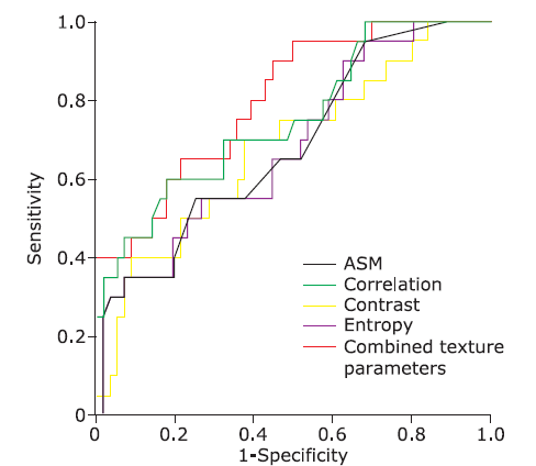
结果 在DWI图像纹理特征参数中,两组间的角二阶矩、对比度、自相关及熵的差异具有统计学意义(P角二阶矩 = 0.014,P对比度 = 0.019,P自相关 = 0.010,P熵 = 0.007)。以上4个指标的ROC曲线下面积为0.685,0.681,0.754和0.683,纳入Logistic回归模型的联合变量(角二阶矩、对比度和熵)的ROC曲线下面积为0.802。二元Logistic回归分析提示角二阶矩、对比度和熵可以作为鉴别乳腺良、恶性肿瘤的变量。

结论 MRI的DWI图像纹理特征分析可以作为乳腺良、恶性肿瘤的鉴别诊断工具。

关键词: 乳腺肿瘤, 纹理分析, 磁共振成像, 鉴别诊断

Abstract:

Objective To investigate the difference in texture features on diffusion weighted imaging (DWI) images between breast benign and malignant tumors.

Methods Patients including 56 with mass-like breast cancer, 16 with breast fibroadenoma, and 4 with intraductal papilloma of breast treated in the Hainan Hospital of Chinese PLA General Hospital were retrospectively enrolled in this study, and allocated to the benign group (20 patients) and the malignant group (56 patients) according to the post-surgically pathological results. Texture analysis was performed on axial DWI images, and five characteristic parameters including Angular Second Moment (ASM), Contrast, Correlation, Inverse Difference Moment (IDM), and Entropy were calculated. Independent sample t-test and Mann-Whitney U test were performed for intergroup comparison. Regression model was established by using Binary Logistic regression analysis, and receiver operating characteristic curve (ROC) analysis was carried out to evaluate the diagnostic efficiency.

Results The texture features ASM, Contrast, Correlation and Entropy showed significant differences between the benign and malignant breast tumor groups (PASM=0.014, Pcontrast=0.019, Pcorrelation=0.010, Pentropy=0.007). The area under the ROC curve was 0.685, 0.681, 0.754, and 0.683 respectively for the positive texture variables mentioned above, and that for the combined variables (ASM, Contrast, and Entropy) was 0.802 in the model of Logistic regression. Binary Logistic regression analysis demonstrated that ASM, Contrast and Entropy were considered as the specific imaging variables for the differential diagnosis of breast benign and malignant tumors.

Conclusion The texture analysis of DWI may be a simple and effective tool in the differential diagnosis between breast benign and malignant tumors.

Key words: breast tumor, texture analysis, magnetic resonance imaging, differential diagnosis

Copyright © 2018 Chinese Academy of Medical Sciences. All right reserved.
 
www.cmsj.cams.cn
京公安备110402430088 京ICP备06002729号-1  Powered by Magtech.

Supervised by National Health Commission of the People's Republic of China

9 Dongdan Santiao, Dongcheng district, Beijing, 100730 China

Tel: 86-10-65105897  Fax:86-10-65133074 

E-mail: cmsj@cams.cn  www.cmsj.cams.cn

Copyright © 2018 Chinese Academy of Medical Sciences

All right reserved.

京公安备110402430088  京ICP备06002729号-1