Chinese Medical Sciences Journal ›› 2019, Vol. 34 ›› Issue (2): 120-132.doi: 10.24920/003587
• Review • Previous Articles Next Articles
A Survey on Intelligent Screening for Diabetic Retinopathy
Dai Yulan1, 3, Zhu Chengzhang2, 3, *( ), Shan Xi1, 3, Cheng Zhenzhen1, 3, Zou Beiji1, 3
- 1. School of Computing Science and Engineering
2. School of Literature and Journalism, Central South University, Changsha 410083, China
3. Hunan Province Engineering Technology Research Center of Computer Vision and Intelligent Medical Treatment, Changsha 410083, China
-
Received:2019-03-22Accepted:2019-05-06Published:2019-05-20Online:2019-05-20 -
Contact:Zhu Chengzhang E-mail:anandawork@126.com
|
We survey DR screening from four perspectives: 1) public color fundus image datasets of DR, 2) DR classification and related lesion-extraction approaches, 3) existing computer-aided systems for DR screening, and 4) existing issues, challenges, and research trends. Our goal is to provide insights for future research directions on DR intelligent screening. |
Cite this article
Dai Yulan,Zhu Chengzhang,Shan Xi,Cheng Zhenzhen,Zou Beiji. A Survey on Intelligent Screening for Diabetic Retinopathy[J].Chinese Medical Sciences Journal, 2019, 34(2): 120-132.
share this article
Add to citation manager EndNote|Reference Manager|ProCite|BibTeX|RefWorks
Table 1
Diabetic retinopathy severity and the grading standards"
| disease severity level | Findings |
|---|---|
| Grade 0: Normal | No visible sign of abnormalities |
| Grade 1: Mild-NPDR | Presence of MAs only |
| Grade 2: Moderate-NPDR | More than just MAs but less than severe NPDR |
| Grade 3: Severe - NPDR | Any of the following: > 20 intraretinal HEs Venous beading Intraretinal microvascular abnormalities no signs of PDR |
| Grade 4: PDR | Either or both of the following: NV Vitreous/pre-retinal HEs |
Table 1
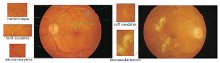
Figure 1.
Manifestations on color fundus images reflecting different pathologies associated with diabetic retinopathy."
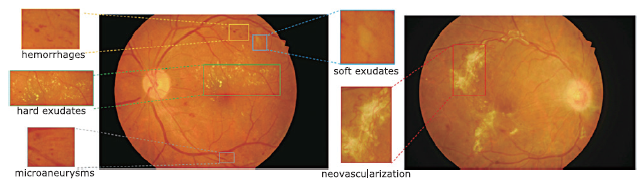
Figure 1.
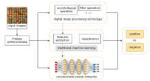
Figure 2.
Technical roadmap of existing methods for diabetic retinopathy detection."

Figure 2.
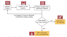
Figure 3.
The general workflow of the intelligent screening system of IDx-DR."

Figure 3.
| [1] |
Yau JWY, Rogers SL, Kawasaki R , et al. Global preva-lence and major risk factors of diabetic retinopathy. Diabetes Care 2012; 35(3):556-64.doi:
doi: 10.2337/dc11-1909 |
| [2] |
Xu Y, Wang L, He J , et al. Prevalence and control of diabetes in Chinese adults. JAMA 2013; 310(9):948-59. doi:
doi: 10.1001/jama.2013.168118 |
| [3] | Zhao KX, Yang PZ, Zhai J , et al. Retinopathy. In: Zhao KX, Yang PZ, editors. Ophthalmology, 8th ed. Beijing: People’s Medical Publishing House; 2013. p. 217-9. |
| [4] |
Kohner EM, Stratton IM, Aldington SJ , et al. Micro-aneurysms in the development of diabetic retinopathy (UKPDS 42). Diabetologia 1999; 42(9):1107-12. doi:
doi: 10.1007/s001250051278 |
| [5] |
Teng T, Lefley M, Claremont D . Progress towards automated diabetic ocular screening: A review of image analysis and intelligent systems for diabetic retinopathy. Med Biol Eng Comput 2002; 40(1):2-13. doi:
doi: 10.1007/BF02347689 |
| [6] |
Hatanaka Y, Nakagawa T, Hayashi Y , et al. CAD scheme to detect hemorrhages and exudates in ocular fundus images. Proceedings of the SPIE of Medical Imaging. 2007 Mar 30; San Diego, USA. 2007. doi: .
doi: 10.1117/12.708367 |
| [7] |
Agurto C, Murray V, Barriga E , et al. Multiscale AM-FM methods for diabetic retinopathy lesion detection. IEEE Trans Med Imaging 2010; 29(2):502-12. doi:
doi: 10.1109/TMI.2009.2037146 |
| [8] |
Ravishankar S, Jain A, Mittal A. Automated feature extraction for early detection of diabetic retinopathy in fundus images. Proceedings of the 22nd IEEE Conference on Computer Vision and Pattern Recognition. 2009 Jun 20-25; Miami, USA. Los Alamitos: IEEE Computer Society Press; 2009. p. 210-7. doi: .
doi: 10.1109/CVPR.2009.5206763 |
| [9] |
Tǎlu S, Cǎlugǎru DM, Lupascu CA . Characterisation of human non-proliferative diabetic retinopathy using the fractal analysis. Int J ophthalmol 2015; 8(4):770-6. doi: .
doi: 10.3980/j.issn.2222-3959.2015.04.23 |
| [10] | Haloi M. Improved microaneurysm detection using deep neural networks. Comput Sci 2015; arXiv: 1505. 04424. |
| [11] |
Prenta?i? P, Lon?ari? S. Detection of exudates in fundus photographs using convolutional neural networks. Proceedings of the 9th IEEE International Symposium on Image and Signal Processing and Analysis. 2015 Sep 7-9; Zagreb, Croatia. Los Alamitos: IEEE Computer Society Press; 2015. p. 188-92. doi: .
doi: 10.1109/ISPA.2015.7306056 |
| [12] |
Tan J H, Fujita H, Sivaprasad S , et al. Automated segmentation of exudates, haemorrhages, microaneurysms using single convolutional neural network. Inf Sci 2017; ( 420):66-76. doi: .
doi: 10.1016/j.ins.2017.08.050 |
| [13] | Kauppi T, Kalesnykiene V, Kamarainen J , et al. DIARETDB0: evaluation database and methodology for diabetic retinopathy algorithms. 2006. |
| [14] |
Kauppi T, Kalesnykiene V, Kamarainen JK , et al. DIARETDB1 diabetic retinopathy database and evaluation protocol. Proceedings of the 11st British Machine Vision Conference. 2007 Sep 10-13; Warwick, UK. 2007. p. 61-5. doi: .
doi: 10.5244/C.21.15 |
| [15] |
Decencière E, Zhang XW, Cazuguel G , et al. Feedback on a publicly distributed image database: The messidor database. Image Anal Stereol 2014; 33(3):231-4. doi:
doi: 10.5566/ias.1155 |
| [16] |
Hoover A, Kouznetsova V, Goldbaum M , et al. Locating blood vessels in retinal images by piecewise threshold probing of a matched filter response. IEEE Trans Med Imaging 2000; 19(3):203-10. doi:
doi: 10.1109/42.845178 |
| [17] |
Odstrcilik J, Kolar R, Budai A , et al. Retinal vessel segmentation by improved matched filtering: evaluation on a new high-resolution fundus image database. IET Image Processing 2013; 7(4):373-83. doi:
doi: 10.1049/iet-ipr.2012.0455 |
| [18] |
Prasanna P, Samiksha P, Ravi K , et al. Indian Diabetic Retinopathy Image Dataset (IDRiD): a database for diabetic retinopathy screening research. Data 2018; 3(3):25-32. doi:
doi: 10.3390/data3030025 |
| [19] | Kaggle Inc. Diabetic Retinopathy Detection. . Published 2015; accessed February 28, 2019.https://www.kaggle.com/c/diabetic-retinopathy-detection/data |
| [20] |
Decencière E, Cazuguel G, Zhang X , et al. TeleOphta: machine learning and image processing methods for teleophthalmology. IRBM 2013; 34(2):196-203. doi:
doi: 10.1016/j.irbm.2013.01.010 |
| [21] |
Niemeijer M, Ginneken BV, Cree MJ , et al. Retinopathy online challenge: automatic detection of microaneurysms in digital color fundus photographs. IEEE Trans Med Imaging 2009; 29(1):185-95. doi:
doi: 10.1109/TMI.2009.2033909 |
| [22] |
Wong Y, Lam B, Lee J , et al. Development of an automatic retinal image analysis (ARIA) for screening cerebral small vessel disease in the community. J Neurol Sci 2017; 381:959. doi:
doi: 10.1016/j.jns.2017.08.2700 |
| [23] |
Prentasic P, Loncaric S, Vatavuk Z , et al. Diabetic reti-nopathy image database (DRiDB): a new database for diabetic retinopathy screening programs research. Proceedings of the 8th International Symposium on Image and Signal Processing and Analysis. 2013 Sep 4-6; Trieste, Italy. Los Alamitos: IEEE Computer Society Press; 2013. p. 712-6. doi: .
doi: 10.1109/ISPA.2013.6703830 |
| [24] |
Winder RJ, Morrow PJ, Mcritchie IN , et al. Algorithms for digital image processing in diabetic retinopathy. Comput Med Imaging Graphics 2009; 33(8):608-22. doi:
doi: 10.1016/j.compmedimag.2009.06.003 |
| [25] |
Lay B, Baudoin C, Klein JC . Automatic detection of microaneurysms in retinopathy fluoro-angiogram. Proceedings of SPIE-the International Society for Optical Engineering. 1984; 432:165-73. doi:
doi: 10.1117/12.936655 |
| [26] |
Spencer T, Olson JA, Mchardy KC , et al. An image-processing strategy for the segmentation and quantification of microaneurysms in fluorescein angiograms of the ocular fundus. Comput Biomed Res Int J 1996; 29(4):284-302. doi:
doi: 10.1006/cbmr.1996.0021 |
| [27] |
Hipwell JH, Strachan F, Olson JA , et al. Automated detection of microaneurysms in digital red‐free photographs: a diabetic retinopathy screening tool. Diabetic Med 2001; 17(8):588-94. doi:
doi: 10.1046/j.1464-5491.2000.00338.x |
| [28] |
Fleming AD, Philip S, Goatman KA , et al. Automated microaneurysm detection using local contrast normalization and local vessel detection. IEEE Trans Med Imaging 2006; 25(9):1223-32. doi:
doi: 10.1109/TMI.2006.879953 |
| [29] |
Puranik SS, Malode VB. Morphology based approach for microaneurysm detection from retinal image. 2016 Processing of the International Conference on Automatic Control and Dynamic Optimization Techniques. 2016 Sep 9-10; Pune, India. Los Alamitos: IEEE Computer Society Press; 2016. p. 635-9. doi: .
doi: 10.1109/ICACDOT.2016.7877663 |
| [30] |
Kamble R, Kokare M. Detection of microaneurysm using local rank transform in color fundus images. ICIP 2017: Processing of the 24th IEEE International Conference on Image Processing. 2017 Sep 17-20; Beijing, China. Los Alamitos: IEEE Computer Society Press; 2017. p. 4442-6. doi: .
doi: 10.1109/ICIP.2017.8297122 |
| [31] |
Gwénolé Q, Lamard M, Josselin PM , et al. Optimal wavelet transform for the detection of microaneurysms in retina photographs. IEEE Trans Med Imaging 2008; 27(9):1230-41. doi:
doi: 10.1109/TMI.2008.920619 |
| [32] |
Zhang B, Wu X, You J , et al. Detection of microaneurysms using multi-scale correlation coefficients. Pattern Recognition 2010; 43(6):2237-48. doi:
doi: 10.1016/j.patcog.2009.12.017 |
| [33] |
Antal B, Hajdu A . An ensemble-based system for microaneurysm detection and diabetic retinopathy grading. IEEE Trans Biomed Eng 2012; 59(6):1720-6. doi:
doi: 10.1109/TBME.2012.2193126 |
| [34] |
Michael A, Ginneken B V, Meindert N , et al. Automatic detection of red lesions in digital color fundus photographs. IEEE Trans Med Imaging 2009; 24(5):584-92. doi:
doi: 10.1109/TMI.2005.843738 |
| [35] |
Dashtbozorg B, Zhang J, Huang F , et al. Retinal microaneurysms detection using local convergence index features. IEEE Trans Image Processing 2018; 27(7):3300-15. doi:
doi: 10.1109/TIP.2018.2815345 |
| [36] |
Wang S, Tang HL, Turk LIA , et al. Localizing microaneurysms in fundus images through singular spectrum analysis. IEEE Trans Biomed Eng 2016; 64(5):990-1002. doi: .
doi: 10.1109/TBME.2016.2585344 |
| [37] |
Xu J, Zhang X, Chen H , et al. Automatic analysis of microaneurysms turn over to diagnose the progression of diabetic retinopathy. IEEE Access 2018; 6:9632-42. doi:
doi: 10.1109/ACCESS.2018.2808160 |
| [38] |
Chudzik P, Majumdar S, Calivá F , et al. Microaneurysm detection using fully convolutional neural networks. Comput Methods Programs Biomed 2018; 158:185-92. doi:
doi: 10.1016/j.cmpb.2018.02.016 |
| [39] |
Budak U, ?engür A, Guo Y , et al. A novel microaneurysms detection approach based on convolutional neural networks with reinforcement sample learning algorithm. Health Inf Sci Syst 2017; 5(1):14. doi:
doi: 10.1007/s13755-017-0034-9 |
| [40] |
Dai L, Fang R, Li H , et al. Clinical report guided retinal microaneurysm detection with multi-sieving deep learning. IEEE Trans Med Imaging 2018: 1149-61. doi: .
doi: 10.1109/TMI.2018.2794988 |
| [41] |
Bae JP, Kim KG, Kang HC , et al. A study on hemorrhage detection using hybrid method in fundus images. J Digital Imaging 2011; 24(3):394-404. doi:
doi: 10.1007/s10278-010-9274-9 |
| [42] |
Xiao D, Yu S, Vignarajan J , et al. Retinal hemorrhage detection by rule-based and machine learning approach. 2017 39th Annual International Conference of the IEEE Engineering in Medicine and Biology Society. 2017 Jul 11-15; Seogwipo, South Korea. Los Alamitos: IEEE Computer Society Press; 2017. p. 660-3. doi: .
doi: 10.1109/EMBC.2017.8036911 |
| [43] |
Tang L, Niemeijer M, Reinhardt JM , et al. Splat feature classification with application to retinal hemorrhage detection in fundus images. IEEE Trans Med Imaging 2013; 32(2):364-75. doi:
doi: 10.1109/TMI.2012.2227119 |
| [44] | Inbarathi R, Karthikeyan R . Detection of retinal hemo-rrhage in fundus images by classifying the splat features using SVM. Int J Innovat Res Sci Eng Technol 2014; 3(3):1979-86. |
| [45] |
Van Grinsven M, Van Ginneken B, Hoyng C , et al. Fast convolutional neural network training using selective data sampling: Application to hemorrhage detection in color fundus images. IEEE Trans Med Imaging 2016; 35(5):1273-84. doi:
doi: 10.1109/TMI.2016.2526689 |
| [46] |
Kande GB, Savithri TS, Subbaiah PV . Automatic detection of microaneurysms and hemorrhages in digital fundus images. J Digital Imaging 2010; 23(4):430-7. doi:
doi: 10.1007/s10278-009-9246-0 |
| [47] |
Akram UM, Khan SA . Automated detection of dark and bright lesions in retinal images for early detection of diabetic retinopathy. J Med Syst 2012; 36(5):3151-62. doi:
doi: 10.1007/s10916-011-9802-2 |
| [48] |
Michael A, Ginneken BV, Meindert N , et al. Automatic detection of red lesions in digital color fundus photographs. IEEE Trans Med Imaging 2005; 24(5):584-92. doi:
doi: 10.1109/TMI.2005.843738 |
| [49] |
Seoud L, Hurtut T, Chelbi J , et al. Red lesion detection using dynamic shape features for diabetic retinopathy screening. IEEE Trans Med Imaging 2016; 35(4):1116-26. doi:
doi: 10.1109/TMI.2015.2509785 |
| [50] |
Adal KM, Van Etten PG, Martinez JP , et al. An automated system for the detection and classification of retinal changes due to red lesions in longitudinal fundus images. IEEE Trans Biomed Eng 2017; 65(6):1382-90. doi: .
doi: 10.1109/TBME.2017.2752701 |
| [51] |
Walter T, Klein JC, Massin P , et al. A contribution of image processing to the diagnosis of diabetic retino-pathy-Detection of exudates in color fundus images of the human retina. Trans Med Imaging 2002; 21(10):1236. doi:
doi: 10.1109/TMI.2002.806290 |
| [52] |
Figueiredo IN, Kumar S, Oliveira CM , et al. Automated lesion detectors in retinal fundus images. Comput Biol Med 2015; 66(C):47-65. doi:
doi: 10.1016/j.compbiomed.2015.08.008 |
| [53] |
Sopharak A, Uyyanonvara B, Barman S , et al. Automatic detection of diabetic retinopathy exudates from non-dilated retinal images using mathematical morphology methods. Comput Med Imaging Graph 2008; 32(8):720-7. doi:
doi: 10.1016/j.compmedimag.2008.08.009 |
| [54] |
Kar SS, Maity S . Automatic detection of retinal lesions for screening of diabetic retinopathy. IEEE Trans Biomed Eng 2018; 65(3):608-18. doi:
doi: 10.1109/TBME.2017.2707578 |
| [55] |
Sánchez CI, Hornero R, López MI , et al. A novel automatic image processing algorithm for detection of hard exudates based on retinal image analysis. Med Eng Physics 2008; 30(3):350-7. doi:
doi: 10.1016/j.medengphy.2007.04.010 |
| [56] |
Li H, Chutatape O . Automated feature extraction in color retinal images by a model based approach. IEEE Trans Biomed Eng 2004; 51(2):246-54. doi:
doi: 10.1109/TBME.2003.820400 |
| [57] |
Wisaeng K, Sa-Ngiamvibool W . Exudates detection using morphology mean shift algorithm in retinal images. IEEE Access 2019; 7:11946-58. doi: 10.1109/ACCESS.2018.2890426.
doi: 10.1109/ACCESS.2018.2890426 |
| [58] |
Youssef D, Solouma NH . Accurate detection of blood vessels improves the detection of exudates in color fundus images. Comput Methods Programs Biomed 2012; 108(3):1052-61. doi:
doi: 10.1016/j.cmpb.2012.06.006 |
| [59] |
Osareh A, Mirmehdi M, Thomas B , et al. Comparative exudate classification using support vector machines and neural networks. in: Dohi T, Kikinis R, editors. Medical Image Computing and Computer Assisted Intervention-MICCAI 2002. Berlin: Springer; 2002. p. 413-20. doi: .
doi: 10.1007/3-540-45787-9_52 |
| [60] |
Osareh A, Shadgar B, Markham R . A computational intelligence-based approach for detection of exudates in diabetic retinopathy images. IEEE Trans Inf Technol Biomed 2009; 13(4):535-45. doi:
doi: 10.1109/TITB.2008.2007493 |
| [61] |
Zhang XW, Thibault G, Decencière E , et al. Exudate detection in color retinal images for mass screening of diabetic retinopathy. Med Image Anal 2014; 18(7):1026-43. doi:
doi: 10.1016/j.media.2014.05.004 |
| [62] |
Harangi B, Antal B, Hajdu A. Automatic exudate detection with improved Na?ve-bayes classifier. 2012 25th IEEE International Symposium on Computer-Based Medical Systems(CBMS). 2012 Jun 20-22; Rome, Italy. Los Alamitos: IEEE Computer Society Press; 2012. p. 139-142. doi: .
doi: 10.1109/CBMS.2012.6266341 |
| [63] |
García M, Sánchez CI, López MI , et al. Neural network based detection of hard exudates in retinal images. Comput Methods Programs Biomed 2009; 93(1):9-19. doi:
doi: 10.1016/j.cmpb.2008.07.006 |
| [64] |
Yu S, Xiao D, Kanagasingam Y. Exudate detection for diabetic retinopathy with convolutional neural networks. 2017 39th Annual International Conference of the IEEE Engineering in Medicine and Biology Society (CMBC). 2017 Jul 11-15; Seogwipo, South Korea. Los Alamitos: IEEE Computer Society Press; 2017. p. 1744-7. doi: .
doi: 10.1109/EMBC.2017.8037180 |
| [65] |
Prenta?i? P, Lon?ari? S . Detection of exudates in fundus photographs using deep neural networks and anatomical landmark detection fusion. Comput Methods Programs Biomed 2016; 137:281-92. doi:
doi: 10.1016/j.cmpb.2016.09.018 |
| [66] |
Herbert FJ, Michael JC, Jorge JGL , et al. Automated segmentation of retinal blood vessels and identification of proliferative diabetic retinopathy. J Opt Soc Am A 2007; 24(5):1448-56. doi:
doi: 10.1364/JOSAA.24.001448 |
| [67] |
Welikala RA, Dehmeshki J, Hoppe A , et al. Automated detection of proliferative diabetic retinopathy using a modified line operator and dual classification. Comput Methods Programs Biomed 2014; 114(3):247-61. doi:
doi: 10.1016/j.cmpb.2014.02.010 |
| [68] |
Lee J, Zee B C, Li Q . Detection of neovascularization based on fractal and texture analysis with interaction effects in diabetic retinopathy. Plos One 2013; 8(12):e75699. doi:
doi: 10.1371/journal.pone.0075699 |
| [69] |
Yu S, Xiao D, Kanagasingam Y . Machine learning based automatic neovascularization detection on optic disc region. IEEE J Biomed Health Inform 2018; 22(3):886-94. doi:
doi: 10.1109/JBHI.2017.2710201 |
| [70] |
Roychowdhury S, Koozekanani DD, Parhi KK , et al. Automated detection of neovascularization for proliferative diabetic retinopathy screening. 2016 38th Annual International Conference of the IEEE Engineering in Medicine and Biology Society (EMBC). 2016 Aug 16-20; Orlando, USA. Los Alamitos: IEEE Computer Society Press; 2016. p. 1300-3. doi: .
doi: 10.1109/EMBC.2016.7590945 |
| [71] |
Goatman KA, Fleming AD, Philip S , et al. Detection of new vessels on the optic disc using retinal photographs. IEEE Trans Med Imaging 2011; 30(4):972-9. doi:
doi: 10.1109/TMI.2010.2099236 |
| [72] |
Nayak J, Bhat PS, Rajendra AU , et al. Automated identification of diabetic retinopathy stages using digital fundus images. J Med Syst 2008; 32(2):107-15. doi:
doi: 10.1007/s10916-007-9113-9 |
| [73] |
Frame AJ, Undrill PE, Olson JA , et al. Texture analysis of retinal neovascularization. Proceedings of the 7th International Conference on Pattern Recognition. 1997 Feb 26-26; London, UK. Los Alamitos: IEEE Computer Society Press; 1997. p. 511-6. doi: .
doi: 10.1049/ic:19970128 |
| [74] |
Pujitha AK, Jahnavi GS, Sivaswamy J. Detection of neovascularization in retinal images using semi-supervised learning. 2017 14th IEEE International Symposium on Biomedical Imaging (ISBI). 2017 Apr 18-21; Melbourne, Australia. Los Alamitos: IEEE Computer Society Press; 2017. p. 688-91. doi: .
doi: 10.1109/ISBI.2017.7950613 |
| [75] |
Gupta G, Kulasekaran S, Ram K , et al. Local characterization of neovascularization and identification of proliferative diabetic retinopathy in retinal fundus images. Comput Med Imaging Graphics 2017; 124-32. doi:
doi: 10.1016/j.compmedimag.2016.08.005 |
| [76] |
Morales S, Engan K, Naranjo V , et al. Retinal disease screening through local binary patterns. IEEE J Biomed Health Inform 2017; 21(1):184-92. doi:
doi: 10.1109/JBHI.2015.2490798 |
| [77] |
Mookiah MRK, Acharya UR, Martis RJ , et al. Evolutionary algorithm based classifier parameter tuning for automatic diabetic retinopathy grading: A hybrid feature extraction approach. Knowledge-Based Syst 2013; 39:9-22. doi:
doi: 10.1016/j.knosys.2012.09.008 |
| [78] |
Rubini SS, Kunthavai A . Diabetic retinopathy detection based on eigenvalues of the hessian matrix. Procedia Comput Sci 2015; 47:311-8. doi:
doi: 10.1016/j.procs.2015.04.001 |
| [79] |
Bhatkar AP, Kharat GU. Detection of diabetic retinopathy in retinal images using MLP classifier. 2015 15th IEEE International Symposium on Nanoelectronic and Information Systems. 2015 Dec 21-23; Indore, India. Los Alamitos: IEEE Computer Society Press; 2015. p. 331-5. doi: .
doi: 10.1109/iNIS.2015.30 |
| [80] |
Gardner G, Keating D, Williamson T , et al. Automatic detection of diabetic retinopathy using an artificial neural network: a screening tool. Br J Ophthalmol 1996; 80(11):940-4. doi:
doi: 10.1136/bjo.80.11.940 |
| [81] |
Usher D, Dumskyj M, Himaga M , et al. Automated detection of diabetic retinopathy in digital retinal images: a tool for diabetic retinopathy screening. Diabetic Med 2004; 21(1):84-90. doi:
doi: 10.1046/j.1464-5491.2003.01085.x |
| [82] |
Szegedy C, Liu W, Jia Y , et al. Going deeper with convolutions. 2015 CVPR: Proceedings of the 28th IEEE Conference Computer Vision and Pattern Recognition. 2015 Jun 7-12; Boston, USA. Los Alamitos: IEEE Computer Society Press; 2015. p. 1-9. doi: .
doi: 10.1109/CVPR.2015.7298594 |
| [83] |
Gulshan V, Peng L, Coram M , et al. Development and validation of a deep learning algorithm for detection of diabetic retinopathy in retinal fundus photographs. J Am Med Assoc 2016; 316(22):2402-10. doi:
doi: 10.1001/jama.2016.17216 |
| [84] |
Quellec G, Charrière K, Boudi Y , et al. Deep image mining for diabetic retinopathy screening. Med Image Anal 2017; 39:178-93. doi:
doi: 10.1016/j.media.2017.04.012 |
| [85] |
Yang Y, Li T, Li W , et al. Lesion detection and grading of diabetic retinopathy via two-stages deep convolutional neural networks. 2017 20th International Conference on Medical Image Computing and Computer-Assisted Intervention (MICCAI). 2017 Sep 10-14; Quebec, Canada. Berlin: Springer; 2017. p. 533-40. doi: .
doi: 10.1007/978-3-319-66179-7_61 |
| [86] |
Ardiyanto I, Nugroho HA, Rlb B. Deep learning-based diabetic retinopathy assessment on embedded system. 2017 39th Annual International Conference of the IEEE Engineering in Medicine and Biology Society (CMBC). 2017, Jul 11-15; Seogwipo, South Korea. Los Alamitos: IEEE Computer Society Press; 2017. p. 1760-3. doi: .
doi: 10.1109/EMBC.2017.8037184 |
| [87] |
Khojasteh P, Aliahmad B, Sridhar PA , et al. Introducing a novel layer in convolutional neural network for automatic identification of diabetic retinopathy. 2018 40th International Conference of the IEEE Engineering in Medicine and Biology Society (CMBC). 2018 Jul 18-21; Honolulu, USA. Los Alamitos: IEEE Computer Society Press; 2018. p. 5938-41. doi: .
doi: 10.1109/EMBC.2018.8513606 |
| [88] |
Gulshan V, Peng L, Coram M , et al. Development and validation of a deep learning algorithm for detection of diabetic retinopathy in retinal fundus photographs. J Am Med Assoc 2016; 316(22):2402-10. doi:
doi: 10.1001/jama.2016.17216 |
| [89] |
Gao ZT, Jie Li J, Guo JX , et al. Diagnosis of diabetic retinopathy using deep neural networks. IEEE Access 2019; 7:3360-70. doi:
doi: 10.1109/ACCESS.2018.2888639 |
| [90] |
Anonymous. FDA permits marketing of first US test labeled for simultaneous detection of tuberculosis bacteria and resistance to the antibiotic rifampin. Clin Infect Dis 2013; 57(8):i-ii. doi: .
doi: 10.1093/cid/cit548 |
| [91] |
Fauw JD, Ledsam JR, Romera-Paredes B , et al. Clinically applicable deep learning for diagnosis and referral in retinal disease. Nature med 2018; 24(9):1342-50. doi: .
doi: 10.1038/s41591-018-0107-6 |
| [92] | Eyenuk Inc. Receives health Canada approval for EyeArt?, an AI-enabled cloud-based automated diabetic retinopathy screening software. Business wire website. d. Published March 8, 2018; Accessed February 28, 2019. |
| [93] | Remidio Innovative Solutions. Smartphone-based retinal imaging together with artificial intelligence powers automated, sensitive and early detection of retinopathy. CISION PR Newswire Website. . Published March 12, 2018; accessed February 28, 2019. |
| [1] | Kunrong Wu, Shufang Zhang, Ziwan Guan, Xiaoli Li, Rui Li, Ying Yin, Yan Li. Methylenetetrahydrofolate Reductase Gene Polymorphism C677T is Associated with Increased Risk of Coronary Heart Disease in Chinese Type 2 Diabetic Patients [J]. Chinese Medical Sciences Journal, 2021, 36(2): 103-109. |
| [2] | Bin Wu,Jianghua Zhou,Wenxin Wang,Huilin Yang,Meng Xia,Binghong Zhang,Zhigang She,Hongliang Li. Association Analysis of Hyperlipidemia with the 28-Day All-Cause Mortality of COVID-19 in Hospitalized Patients [J]. Chinese Medical Sciences Journal, 2021, 36(1): 17-26. |
| [3] | Honglin Zu,Likun Hou,Hongwei Liu,Yuanbo Zhan,Ju He. Identify Candidate Genes in the Interaction between Abdominal Aortic Aneurysm and Type 2 Diabetes Mellitus by Using Biomedical Discovery Support System [J]. Chinese Medical Sciences Journal, 2021, 36(1): 50-56. |
| [4] | Lin Ye, Wang Zhenlian, Yan Min, Zhu Feiyu, Duan Ye, Sun Zhiqin. Effect of Trimetazidine on Diabetic Patients with Coronary Heart Diseases: A Meta-Analysis of Randomized, Controlled Trials [J]. Chinese Medical Sciences Journal, 2020, 35(3): 226-238. |
| [5] | Shen Chang, Zhao Meng, Li Yunyun, Liu Ningpu. Methylenetrahydrofolate Reductase Gene C677T Polymorphism and Diabetic Retinopathy: a Meta-Analysis [J]. Chinese Medical Sciences Journal, 2020, 35(1): 71-84. |
| [6] | Yin Ying, Li Rui, Li Xiaoli, Wu Kunrong, Li Ling, Xu Yuedong, Liao Lin, Yang Rui, Li Yan. Association Between Homocysteine Level and Methylenetetrahydrofolate Reductase Gene Polymorphisms in Type 2 Diabetes Accompanied by Dyslipidemia [J]. Chinese Medical Sciences Journal, 2020, 35(1): 85-91. |
| [7] | Chen Qiang, Zhang Liwei, Huang Dangsheng, Zhang Chunhong, Wang Qiushuang, Shen Dong, Xiong Minjun, Yang Feifei. Five-year Clinical Outcomes of CAD Patients Complicated with Diabetes after StentBoost-optimized Percutaneous Coronary Intervention [J]. Chinese Medical Sciences Journal, 2019, 34(3): 177-183. |
| [8] | Chen Zhiye, Zang Xiujuan, Liu Mengqi, Liu Mengyu, Li Jinfeng, Gu Zhaoyan, Ma Lin. Abnormal Alterations of Cortical Thickness in 16 Patients with Type 2 Diabetes Mellitus: A Pilot MRI Study△ [J]. Chinese Medical Sciences Journal, 2017, 32(2): 75-82. |
| [9] | Du Junhui, Li Rong. Can Fundus Fluorescein Angiography be Performed for Diabetic Patients on Oral Metformin?△ [J]. Chinese Medical Sciences Journal, 2017, 32(2): 119-122. |
| [10] | Yang Yingying, Pan Hui, Wang Bo, Chen Shi, Zhu Huijuan. Efficacy and Safety of SGLT2 Inhibitors in Patients with Type 1 Diabetes: A Meta-analysis of Randomized Controlled Trials [J]. Chinese Medical Sciences Journal, 2017, 32(1): 22-27. |
| [11] | Jing Yang, Mei-cen Zhou, Kai Feng, Ou Wang, Hua-bing Zhang, Wei Li, Fan Ping, Jing Yang, Yu-xiu Li. Clinical Characteristics of 261 Cases of Hospitalized Patients with Type 1 Diabetes Mellitus [J]. Chinese Medical Sciences Journal, 2016, 31(2): 69-75. |
| [12] | Ying Fan, Shan-xiao Zhang, Meng Ren, Li-feng Hong, Xiao-ni Yan. Impact of 1, 25-(OH)2D3 on Left Ventricular Hypertrophy in Type 2 Diabetic Rats [J]. Chinese Medical Sciences Journal, 2015, 30(2): 114-120. |
| [13] | Yu Deng,Xiu-fen Yang,Hong Gu,Apiradee Lim,Munkhtulga Ulziibat,Torkel Snellingen,Jun Xu,Kai Ma,Ning-pu Liu. Association of C(-106)T Polymorphism in Aldose Reductase Gene with Diabetic Retinopathy in Chinese Patients with Type 2 Diabetes Mellitus [J]. Chinese Medical Sciences Journal, 2014, 29(1): 1-6. |
| [14] | Yi-jun Zhou*,Ai Li, Yu-ling Song, Hui Zhou, Yan Li, Yin-si Tang. Role of Sclerostin in the Bone Loss of Postmenopausal Chinese Women with Type 2 Diabetes [J]. Chinese Medical Sciences Journal, 2013, 28(3): 135-139. |
| [15] | Liu-mei Hu, Xia Lei, Bo Ma, Yu Zhang, Yan Yan, Ya-lanWu, Ge-zhi Xu,Wen Ye, Ling Wang,Guo-xu Xu, Guo-tong Xu*, Wei-ye Li. Erythropoietin Receptor Positive Circulating Progenitor Cells and Endothelial Progenitor Cells in Patients with Different Stages of Diabetic Retinopathy [J]. Chinese Medical Sciences Journal, 2011, 26(2): 69-76. |
| Viewed | ||||||
|
Full text |
|
|||||
|
Abstract |
|
|||||
|